A walk around the block, a few hours of focused work, a social event, or a workout you thought you were finally ready for. It didn’t feel excessive at the time.
Then, 12 to 72 hours later, something happened that has no parallel in “normal” human experience. Not tiredness. Not soreness. A complete systemic collapse, with fatigue so profound it feels neurological, brain fog that makes thinking feel like wading through quicksand, muscle pain, sensory sensitivity, and a return of every symptom you thought you’d been making progress on.
This is post-exertional malaise. And if you have Long COVID or Post-Vaccine Syndrome, you’ve almost certainly experienced it.
What you may not have received is an honest clinical explanation of what it actually is, why it happens, and why the treatment most clinicians prescribe — pushing through, gradually increasing activity, “deconditioning” management — is not just ineffective but genuinely harmful.
At Leading Edge Clinic, we treat PEM as one of the most mechanistically complex and most clinically important features of Long COVID and Post-Vaccine Syndrome. Getting it wrong doesn’t just stall recovery. It actively damages the biology you’re trying to repair.
What Post-Exertional Malaise Actually Is
PEM is formally defined as the worsening of symptoms following physical, mental, or cognitive exertion. It typically occurs with a delay of 12 to 48 hours after the triggering activity, and can last days to weeks.
The delay is important because it’s what makes PEM so consistently misunderstood by clinicians who aren’t familiar with it. If you crashed immediately after exertion, it would feel like an obvious cause-and-effect relationship. The delay means that by the time you feel the worst of it, you’ve already returned to baseline and the connection to what you did two days ago is no longer obvious. Patients are told their symptoms are unrelated to exercise. Sometimes they get an even worse explanation… “it’s all in your head”. Clinicians interpret the crash as a separate event rather than a predictable physiological consequence.
PEM is not deconditioning. It is not anxiety. It is definitely not a failure of effort or will – many of our patients were often “high achievers” prior to their illness. PEM is a specific, reproducible, biologically mediated response to exertion that occurs in a body with compromised cellular energy production, impaired microcirculation, and immune dysregulation. Even the mainstream research now makes this unambiguous.
The Biology: What Is Actually Happening During a PEM Crash
Mitochondrial Dysfunction at the Core
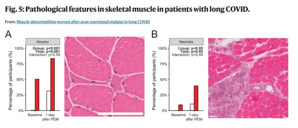
The most important mechanistic development in understanding PEM in Long COVID came from a series of muscle biopsy studies — most notably the 2024 landmark study published in Nature Communications by Appelman, Charlton, and colleagues at Amsterdam UMC, and the accompanying opinion article in Trends in Endocrinology and Metabolism by the same group.
What these studies found in the skeletal muscle tissue of Long COVID patients was as follows: intrinsic mitochondrial dysfunction, not just as a baseline impairment, but one that worsened measurably after exercise. Mitochondrial respiration (the process by which cells produce ATP) decreased significantly one day after maximal, PEM-inducing exercise. Markers of mitochondrial density dropped. The muscle showed acute tissue damage, focal necrosis, and intramuscular infiltration of immune cells that was not present before the exercise challenge.
This is not deconditioning. A deconditioned person’s muscles don’t necrotize after moderate exercise. What’s happening in Long COVID and Post-Vaccine Syndrome PEM is closer to a pathological injury response. The tissue cannot meet the energetic demand placed on it, and the attempt to do so causes measurable cellular damage.
The same research group also identified a shift in muscle fiber type, away from slow-twitch oxidative fibers and toward fast-twitch glycolytic fibers. This matters because oxidative fibers rely on efficient mitochondrial energy production for sustained activity. Glycolytic fibers burn energy quickly but produce lactate rather than sustained ATP. We have heard countless times from patients describing their PEM, some variation of, “it feels like my entire body is full of lactic acid. Like I’ve been poisoned.” A muscle that has shifted toward glycolytic metabolism will hit an energy wall faster, recover more slowly, and be more vulnerable to the kind of anaerobic overload that characterizes PEM.
The Microcirculation Problem
The mitochondrial dysfunction doesn’t occur in isolation. It is compounded by, and in part caused by, the endothelial dysfunction and microclotting that are central features of Long COVID.
When capillary beds are partially obstructed by amyloid fibrin microclots, oxygen delivery to working muscle tissue is impaired. The mitochondria of a Long COVID patient’s muscle cells may be operating in a relative hypoxic environment, even at rest. Under the increased oxygen demand of exercise, that deficit becomes significant. The tissue is asked to produce more energy precisely when its oxygen supply is most constrained.
A 2023 study in Acta Neuropathologica confirmed that Long COVID patients show capillary alterations and immune dysregulation in skeletal muscles. In other words, the physical infrastructure of oxygen delivery to muscle tissue is damaged. You cannot exercise your way out of damaged capillaries. Graded exercise therapy aimed at improving mitochondrial function in these patients is asking an engine with a clogged fuel line to run harder.
The Immune Activation Component
Exercise, even moderate exercise, triggers an inflammatory response in healthy individuals. The muscles release cytokines. Immune cells infiltrate tissue. This is part of normal adaptation and recovery. In healthy individuals, the process resolves and the tissue rebuilds stronger.
In Long COVID and Post-Vaccine Syndrome patients with already-elevated inflammatory signaling (already-elevated IL-6, already-activated mast cells, already-dysregulated immune responses), exercise delivers an additional inflammatory load onto a system with no reserve capacity to manage it. The immune activation triggered by exercise doesn’t resolve normally. It compounds the existing inflammatory state, triggering the systemic symptom cascade that characterizes a PEM crash.
Research published in PMC in 2025 confirmed that PEM in Long COVID is mediated by dysfunctions in both mitochondrial capacity and microcirculation, maintained by latent immune activation that conjointly impairs peripheral bioenergetics. This is not a psychological phenomenon. It is a coordinated biological failure at the cellular and vascular level.
The Cell Danger Response Connection
There is a deeper layer to PEM that connects to the Cell Danger Response framework we’ve written about previously.
When cells are in an active CDR state (as many Long COVID and Post-Vaccine Syndrome patients’ cells are), exertion represents exactly the kind of additional stressor that further activates the danger signaling cascade. ATP is released into the extracellular space as a danger signal. The purinergic signaling system, already dysregulated, interprets the metabolic stress of exercise as renewed threat. Rather than recovering normally, cells double down on the defensive metabolic state, suppressing normal energy production further, perpetuating the CDR, and deepening the very dysfunction that made exercise difficult in the first place.
This mechanism helps explain why PEM can leave patients worse off than before the triggering activity, not just temporarily, but potentially persistently if the CDR activation is severe enough to push cells further into the senescent cascade.
The Graded Exercise Therapy Problem
Here is the clinical position that many uninformed clinicians are still operating from: Long COVID fatigue and exercise intolerance are primarily driven by deconditioning, and the solution is gradual, progressive increases in physical activity. This is also known as Graded exercise therapy, or GET.
This position is not just wrong, it is dangerous. It has been formally disputed by clinicians and researchers on the basis of the biological evidence, it was used in the ME/CFS context based on a deeply flawed trial (the PACE trial), and its application to Long COVID (and PACVS) patients with PEM has been specifically and publicly opposed by leading researchers including those at Amsterdam UMC, Mt. Sinai, and multiple European academic centers.
In January 2024, a letter published in Nature signed by Dutch, German, and Austrian investigators (joined by David Putrino of Mt. Sinai) stated explicitly: “We cannot agree with the recommendations for graded exercise therapy for people living with Long COVID who have post-exertional malaise.” The letter noted that the PACE trial, which is frequently cited in support of GET, has had its results called into question due to substantial protocol deviations and retrospective adjustment of recovery criteria. Improperly interpreted and manipulated study design and results? Where else have we seen that?
Professor Todd Davenport, a physical therapy researcher at the University of the Pacific, stated bluntly in 2024: “Any article published in 2024 citing the PACE trial as evidence of safety and efficacy of graded exercise should not be taken seriously.”
A review of exercise trials in Long COVID published in The Sick Times in late 2025 found that of 112 exercise-related trial registrations for Long COVID, fewer than 20% even mentioned PEM. Of those that did, two excluded participants with moderate-to-severe PEM and two excluded PEM participants entirely. The research community is studying exercise in Long COVID while systematically excluding the patients for whom exercise is most dangerous.
The deconditioning model fails for a simple reason: a deconditioned person’s skeletal muscle does not show focal necrosis, mitochondrial enzyme collapse, and immune cell infiltration after moderate exercise. That is what Long COVID patients’ muscle tissue shows. The biology is not consistent with deconditioning. It is consistent with a pathological injury response in tissue that lacks the cellular and vascular infrastructure to handle exertional demand.
What Appropriate PEM Management Actually Looks Like
If graded exercise therapy is the wrong answer, what is the right one? The honest clinical answer has two parts: understanding your energy envelope, and treating the underlying mechanisms that are causing PEM in the first place.
Pacing and the Energy Envelope
Pacing is not giving up. Many patients struggle to reconcile between their former selves, and their new physical constraints. But, pacing is a clinically rational strategy based on the biology of PEM. Specifically, the observation that there is a physiological threshold below which activity is tolerable and above which it triggers the pathological crash cascade.
The goal of pacing is to identify and stay within your energy envelope. Your energy envelope is the level of exertion your body can sustain without triggering post-exertional symptom worsening. This requires honest, often uncomfortable acceptance that your current energy envelope may be far smaller than your pre-illness capacity. Attempting to expand that envelope by pushing through symptoms (the instinct that works for normal fatigue, and has been socially built into many of our mindsets) actively damages the biology you’re trying to repair. It is easy to understand why patients may feel like they are going to “fall behind”, when adapting to this newfound situation.
Heart rate monitoring can be a useful pacing tool. Research suggests that staying below the anaerobic threshold, often approximated as 50–60% of age-predicted maximum heart rate in Long COVID patients, reduces the probability of triggering a crash. This is a lower threshold than most patients expect and lower than most clinicians prescribe.
Another extremely important and nuanced note… Cognitive and emotional exertion count. PEM is not exclusively triggered by physical activity. Mental effort, social interaction, sensory stimulation, and emotional stress can all trigger crashes in patients with significant PEM. This is consistent with: 1) the neurological and circulating contributors to PEM that the research has identified; and 2) the physiological facts that our brains account for 20% of our daily energy expenditure. All this means that pacing must encompass the full scope of daily exertion, not just physical exercise.
Treating the Underlying Mechanisms
Pacing manages the symptom. It does not treat the cause. For Long COVID and Post-Vaccine Syndrome patients, the cause of PEM is the biological substrate described above: mitochondrial dysfunction, impaired microcirculation, immune dysregulation, and the ongoing CDR state and senescent cell burden.
Addressing microclotting. If capillary obstruction from amyloid fibrin microclots is impeding oxygen delivery to muscle tissue, reducing that obstruction through appropriate fibrinolytic support guided by clinical assessment (and where relevant, PAI-1 genotype) directly addresses one of the key contributors to PEM.
Mitochondrial support. The evidence for mitochondrial dysfunction in PEM is now well-established. Supporting mitochondrial function represents a rational clinical approach, though responses are individual and should be guided by a clinician familiar with this patient population.
Reducing the inflammatory environment. Since immune activation during exercise compounds the existing inflammatory state and drives PEM crashes, reducing the baseline inflammatory burden expands the effective energy envelope over time.
CDR-targeted approaches. For patients where the Cell Danger Response is a significant driver, interventions that support purinergic signaling normalization and allow cells to progress through the healing cycle are directly relevant to PEM. Reducing CDR activation reduces the hair-trigger sensitivity to exertion that characterizes severe PEM.
Careful sequencing. None of these interventions operates in isolation. PEM in Long COVID typically reflects multiple simultaneous mechanisms, and the clinical art is in identifying which mechanisms are dominant in a given patient and addressing them in the right sequence. A patient whose primary PEM driver is microclotting needs a different initial emphasis than a patient whose primary driver is MCAS-mediated immune activation after exercise.
What We Tell Our Patients
At Leading Edge Clinic, the practical guidance we give patients with significant PEM is this:
PEM is a real biological phenomenon with measurable cellular and vascular causes. It is not a symptom you can push through. Attempting to do so, without first addressing the underlying biology that is causing it, will not improve your condition… it will damage it.
However, there is good news. Your current energy envelope is not a permanent ceiling. It is a reflection of your current biological state. As we work on the underlying mechanisms (microclotting, immune dysregulation, CDR, mitochondrial function) your energy envelope will expand. We have seen it over and over. But that expansion has to come from biology improving, not from forcing activity in a system that isn’t ready for it.
This process takes time. It is non-linear. There will be weeks that feel like setbacks. Honest pacing and the discipline to stay within your current envelope even when you feel temporarily better is one of the most important things you can do to protect your recovery trajectory.
Recovery from PEM-dominated Long COVID is one of the slower trajectories we manage, and one of the most satisfying when it happens. Patients who have been bed-bound or housebound for years have regained meaningful function. But that recovery almost always required first stopping the cycle of crash and partial recovery that graded exercise therapy perpetuates, and replacing it with intelligent, mechanistically-informed clinical management.
If you’re interested in receiving care for your PEM, you can register here.
Leading Edge Clinic specializes in Long COVID, Post-Vaccine Syndrome, and complex post-infectious illness. Dr. Pierre Kory, Scott Marsland, FNP-C, and the rest of our clinical team have treated more than 3,500 patients, many who suffer with PEM. We see patients in all 50 states via telehealth.
This article is for educational purposes and does not constitute medical advice.
Key References
Appelman B, Charlton BT, Goulding RP, et al. Muscle abnormalities worsen after post-exertional malaise in Long COVID. Nat Commun. 2024;15:17. https://doi.org/10.1038/s41467-023-44432-3
Charlton BT, Goulding RP, Jaspers RT, et al. Skeletal muscle adaptations and post-exertional malaise in Long COVID. Trends Endocrinol Metab. 2025;36(7):614–622. https://doi.org/10.1016/j.tem.2024.11.008
Aschman T, et al. Post-COVID exercise intolerance is associated with capillary alterations and immune dysregulations in skeletal muscles. Acta Neuropathol Commun. 2023;11:193. https://doi.org/10.1186/s40478-023-01662-2
Towards an understanding of physical activity-induced post-exertional malaise: microvascular alterations and immunometabolic interactions in post-COVID and ME/CFS. PMC. 2025. https://pmc.ncbi.nlm.nih.gov/articles/PMC11825644/
Vink M, Vink-Niese A. CBT and graded exercise therapy studies have proven that ME/CFS and Long COVID are physical diseases. Front Hum Neurosci. 2025. https://pmc.ncbi.nlm.nih.gov/articles/PMC11814198/
Introduction: The Wrong Question About Reinfection
Every time a new COVID variant emerges, the public health conversation follows the same script. Is it more severe? How many mutations does it have? Will the current vaccine protect against it (hah!)? Are hospitalizations rising?
For people with Long COVID, Post-Vaccine Syndrome, or any history of spike protein-related illness, they are almost entirely the wrong questions.
The correct question isn’t whether the COVID, or the latest variant Cicada variant (BA.3.2), causes more severe acute illness than previous strains. The correct question is: what does each additional COVID exposure do to someone who already has persistent spike protein, ongoing cellular senescence, and an immune system that never fully resolved the first round?
The answer, supported by emerging research from scientists at the Vedicinals group and consistent with what we observe clinically, is deeply concerning. It is also largely absent from mainstream COVID coverage.
This post explains why reinfection matters in a way that the standard messaging doesn’t capture, what the Cicada variant means specifically for this patient population, and what can be done to reduce the biological cost of repeated exposure.
What the Cicada Variant Actually Is
As of early April 2026, BA.3.2 has been detected in more than half of US states, according to the CDC. The WHO classified BA.3.2 as a variant under monitoring in December 2025, citing the variant’s many mutations and substantial antibody escape.
What makes Cicada notable is not its acute severity, but its spike protein. Compared to the current predominant strains of SARS-CoV-2, BA.3.2 carries 70 to 75 genetic changes in its spike protein. Of course, we alredy know the spike protein drives fibrinogen misfolding, mast cell activation, endothelial dysfunction, microclotting, and cellular senescence in Long COVID patients. But, Cicada’s mutations may help it evade antibodies. In a patient population where immune dysfunction is already significant, this is something Long COVID and Post-Vaccine Syndrome patients need to take note of for reasons explained later in this post.
Importantly, early data suggests BA.3.2 does not cause more severe acute disease than recent variants.
For the general public, that assessment is probably reassuring. For patients with existing spike protein burden and senescent cell accumulation, it misses the point entirely.
The Framework That Changes Everything: Spike Persistence and the Senescence Cascade
To understand why reinfection matters so much for this patient population, you need to understand a mechanistic framework that is only now beginning to enter the scientific literature, and that we at Leading Edge Clinic have been treating clinically.
A recently published paper from researchers at the Vedicinals group and collaborating institutions advances a hypothesis that brings together two phenomena that have been observed separately but rarely connected with sufficient clinical clarity – persistent spike protein production and progressive cellular senescence.
The key insight is this: Long COVID may not be primarily a problem of “how much spike is left over” from an infection. It may be a problem of ongoing spike production from a small population of cells that never stopped making it.
The Persistent Producer Cell
The paper proposes that a small number of cells, potentially harboring viral RNA in protected intracellular compartments called double-membrane vesicles, or having integrated spike-encoding genetic material, continue to produce and release spike protein long after the acute infection resolves. These “producer cells” generate modest amounts of spike individually, but their output is continuous, and it accumulates in tissues over time.
This would explain a striking observation from a high-volume European Long COVID diagnostic laboratory: spike protein detectability in Long COVID patients rose from roughly 30–40% in 2024, to approximately 75% in the first three quarters of 2025, to 96.5% by the fourth quarter of 2025. Under a simple “leftover antigen slowly clearing” model, you would expect a declining curve. A rising curve points toward ongoing production – an active source term, not residual debris from a resolved infection.
How Spike Spreads Beyond the Original Source
The paper outlines several mechanisms by which spike from a small number of producer cells can reach a vastly larger number of healthy cells:
Extracellular vesicles (exosomes). Spike protein packaged inside exosomes can evade antibody neutralization. Antibodies can’t bind to what they can’t reach. These vesicles carry spike through circulation and into tissues, delivering it to cells that never encountered the virus directly. These are the same vesicles by which proposed spike protein shedding events occur.
Syncytia formation. When spike-expressing cells contact cells with ACE2 receptors, they can fuse, creating multinucleated structures. Each fusion event effectively transfers spike-producing capacity to multiple adjacent cells simultaneously. What does this mean? It means a single producer cell can transfect 5–20 neighbors in one fusion event.
Tunneling nanotubes (TNTs). Cells can form thin, direct membrane bridges to neighboring cells through which spike protein, vesicles, and potentially viral RNA can transit — entirely shielded from antibody neutralization. A spike-producing cell may maintain TNT connections with 5–50 neighboring cells at once.
The result is progressive tissue saturation: a small upstream source driving a disproportionately large downstream burden.
The Senescence Cascade
The senescence cascade is where the model becomes clinically relevant.
As cells accumulate spike protein intracellularly – whether through exosomal uptake, syncytia fusion, or TNT transfer – they experience proteostatic stress, ER stress, and DNA-damage-response signaling. Once these signals cross a threshold, the cell enters a state of irreversible growth arrest: cellular senescence. It stops dividing, resists programmed cell death, and begins producing a continuous stream of pro-inflammatory signals collectively called the SASP — the senescence-associated secretory phenotype.
But the most important feature of senescent cells is that their SASP is contagious.
SASP factors — IL-6, IL-8, IL-1β, TNF-family molecules, matrix metalloproteinases — induce senescence in neighboring cells that may contain no measurable spike antigen themselves. This bystander or paracrine senescence means the lesion expands far beyond the originally spike-exposed population. Cells that never encountered spike become senescent because they were next to cells that did.
This creates a self-amplifying cascade that the paper describes precisely: the disease can transition from a spike-driven initiation phase to a senescence-dominant maintenance phase. In this case, the symptom burden becomes partially decoupled from measurable spike load. Patients remain severely symptomatic even when standard tests don’t detect viral material, because the biology has “handed off” from an antigenic driver to a self-sustaining cellular program.
This is consistent with what we observe clinically in Long COVID patients who have been ill for two, three, or four years. It is also consistent with why therapies aimed purely at viral clearance often underperform in established disease.
Why Each Reinfection Layers Onto This Foundation
Each additional COVID exposure, whether from Cicada, any other variant, or a future strain, adds a new round of spike protein input to a system that is already struggling with ongoing production, progressive tissue saturation, and expanding senescent cell burden.
It replenishes the upstream source. A reinfection doesn’t just cause acute illness and then clear. It potentially seeds new producer cells, adds to the extracellular vesicle pool carrying spike, and re-exposes tissues that may have been recovering toward a threshold. The spike positivity data from the Vedicinals group’s European laboratory is consistent with this: a population of Long COVID patients showing rising, not declining, spike detection over time. Reinfections accelerate what was already an accumulating burden.
It compounds the senescence burden. Each new wave of spike-driven senescence induction is additive. Primary senescence from new spike exposure layers onto existing senescent cell populations. The paracrine cascade expands into new tissue territory. Patients who were at the margin of clinical stability (still functioning, managing their condition) can cross a tipping point following reinfection into significantly more impaired states.
The Cicada variant’s immune escape makes it more likely to establish a productive reservoir. For a Long COVID patient whose immune system is already dysregulated and whose neutralizing antibody response may be quantitatively or qualitatively impaired, a variant with enhanced antibody escape has a higher probability of establishing persistent producer cells rather than being rapidly cleared.
The spike protein variant may matter. The Vedicinals paper notes that earlier variants, including Omicron, appear to induce higher p16 and p21 expression (markers of cellular senescence) than ancestral strains, despite causing less severe acute illness. Cicada’s 70–75 spike mutations represent a distinct protein configuration. Whether this configuration affects senescence-induction kinetics is not yet established, but the direction of the existing data (that newer variants may be more senescence-inducing despite less acute severity) is something this patient population needs to take seriously.
The acute illness is not the danger. The downstream biology is.
This is the core message that is absent from every piece of mainstream Cicada variant coverage. The question “is it more severe?” is answered by hospitalization rates and ICU admissions. It does not capture what happens over the following 6, 12, or 18 months in someone already carrying significant senescent cell burden. It does not capture the cumulative cost of each reinfection to a biological system that has been running in a state of chronic dysregulation.
The Role of Senolytics: Reducing the Cost of Repeat Exposure
If the senescence cascade model is correct, and the evidence is increasingly consistent with it, then one of the most important things a Long COVID patient can do in the context of ongoing variant circulation is actively work to reduce their existing senescent cell burden. This reduces the foundation onto which a new exposure would layer, and it may limit the propagation of any new senescence cascade.
This is not a new clinical concept for us. Senolytic interventions (agents that selectively clear senescent cells) have been part of our treatment framework for Long COVID and Post-Vaccine Syndrome.
Some (but not all) clinically relevant therapies in this context include:
Intermittent fasting and autophagy promotion. The body’s cellular recycling program, autophagy, is one of the primary endogenous mechanisms for clearing dysfunctional cellular components. Caloric restriction and intermittent fasting protocols can meaningfully upregulate autophagy, and this can complement other senolytic approaches. However, as we’ve noted in our MCAS content, fasting protocols in Long COVID patients require clinical judgment. Not every patient can tolerate aggressive fasting, and the approach needs to be sequenced appropriately with other interventions.
Senomorphics — reducing SASP without clearing cells. For patients where aggressive senolytic dosing is not appropriate, senomorphic agents — those that reduce SASP output without necessarily clearing the senescent cells — can limit the paracrine propagation of the cascade. Low-dose rapamycin and metformin both have evidence in this area, and both are already used in relevant clinical contexts at our practice.
A 2021 study published in Science by Camell and colleagues provided direct evidence that senolytics reduce coronavirus-related mortality in aged mice, specifically by clearing the senescent cell burden that amplified the inflammatory response to viral infection. While this was in the context of acute infection rather than chronic Long COVID, the mechanism is directly relevant. Reducing pre-existing senescent cell burden before or after a new exposure limits how much the SASP-driven inflammatory cascade can amplify in response.
This is a clinically actionable implication. Patients with established Long COVID who maintain an ongoing senolytic protocol are not only treating their current disease, they are reducing the biological cost of the next inevitable exposure.
What This Means Practically for Long COVID and PACVS Patients Right Now
The COVID variant of the week is circulating in a population of Long COVID patients who are already carrying spike protein burden, established senescent cell populations, and a SASP-driven inflammatory environment.
The practical implications for this population are:
Reinfection prevention matters more for you than for the general population. Your risk includes compounding senescence, new producer cell seeding, potential tipping-point transitions in disease severity.
Senolytic maintenance is relevant timing. For patients already on senolytic protocols, ensuring adequate maintenance dosing during a period of active variant circulation is clinically sensible. For patients who have not yet incorporated senolytics into their treatment, this is a reasonable moment to discuss it with your provider.
Acute COVID treatment matters differently for you. If you do develop an acute Cicada infection, early intervention with antiviral therapy is relevant not primarily to prevent severe acute illness, but to limit the duration and magnitude of spike protein production, and therefore the new producer cell burden established during the infection. Shorter, lower-severity infection means less spike, means less new senescence induction.
The spike protein clearance and senolytic work you do now is investment against future exposures. Reducing your current spike burden and your current senescent cell load is not just about feeling better today, it is about building a lower biological baseline from which any future reinfection would cascade. This is why we treat Long COVID not as a single-point intervention but as an ongoing, evolving clinical relationship.
Conclusion: The Real Risk of Reinfection
The Cicada variant is being reported as “not more severe”. For the acute phase, that is accurate. But for the Long COVID and Post-Vaccine Syndrome patient, the frame of acute severity is inadequate.
The real risk of reinfection is not hospitalization. The real risk is what each additional exposure does to a biological system already operating under chronic spike pressure and expanding senescent cell burden. Each reinfection is not a reset. It is an addition to a running total that has consequences that unfold over months and years.
The senescence cascade hypothesis advanced by researchers at the Vedicinals group provides a mechanistic vocabulary for why patients experience progressive worsening over time despite no acute event. It explains why Long COVID symptoms can outlast any measurable marker of infection. And it clarifies what is at stake in the context of ongoing variant circulation.
This is the conversation that needs to be happening with Long COVID patients right now. At Leading Edge Clinic, it is the conversation we are having.
Leading Edge Clinic specializes in Long COVID, Post-Vaccine Syndrome, and complex post-infectious illness. Our providers treat patients across all 50 states via telehealth.
This article is for educational purposes and does not constitute medical advice.
Key References
Gerlach J, Baig AM, et al. Persistent Spike Protein Production and Progressive Tissue Saturation in Long COVID: Novel Hypothesis for a Senescence Cascade. Vedicinals Group / Health-Shield. 2025. [Preprint]
Camell CD, Yousefzadeh MJ, Zhu Y, et al. Senolytics reduce coronavirus-related mortality in old mice. Science. 2021;373:eabe4832. https://doi.org/10.1126/science.abe4832
Patterson BK, et al. Persistence of SARS-CoV-2 S1 protein in CD16+ monocytes in PASC up to 15 months post-infection. Front Immunol. 2021;12:746021. https://doi.org/10.3389/fimmu.2021.746021
Meyer K, et al. SARS-CoV-2 spike protein induces paracrine senescence and leukocyte adhesion in endothelial cells. J Virol. 2021;95:e00794-21. https://doi.org/10.1128/JVI.00794-21
Tsuji S, et al. SARS-CoV-2 infection triggers paracrine senescence and a sustained senescence-associated inflammatory response. Nat Aging. 2022. https://doi.org/10.1038/s43587-022-00170-7
Acosta JC, et al. A complex secretory program orchestrated by the inflammasome controls paracrine senescence. Nat Cell Biol. 2013;15(12):1524–1535. https://doi.org/10.1038/ncb2871
Introduction: The Clotting Problem Most Doctors Aren’t Testing For
Many Long COVID and Post-Vaccine Syndrome patients know their symptoms. Fatigue that doesn’t resolve with rest. Brain fog that feels like you’re wading through muck just to formulate a coherent thought. Breathlessness that appears without warning. Chest tightness that comes and goes. Post-exertional crashes that wipe out any attempt at normal activity.
What most patients, and most clinicians, have come to know is that for a significant proportion of these patients, the blood itself is part of the problem.
Not in the dramatic, visible way that shows up on imaging. In a quieter, more insidious way: tiny, abnormal clots forming throughout the microvasculature, reducing oxygen delivery to tissues, trapping inflammatory molecules, and physically obstructing circulation in capillaries too small to show up on any standard scan. Amyloid fibrin microclots are not a novel issue. But, with spike protein, they are more significant than ever before. Not a single patient of ours comes to us with low levels of microclotting.
A critical aspect of microclotting we will explore in this article: a genetic variant that approximately 25–30% of the general population carries means some patients are dramatically more prone to forming these clots, and dramatically less able to clear them, than others.
Understanding this mechanism, and how your genetics interact with it, changes what treatment should look like. At Leading Edge Clinic, it’s something we assess in patients where microclotting is suspected as a significant driver. This post explains the underlying science and the clinical implications.
What Are Fibrin Amyloid Microclots?
Normal blood clotting is a tightly regulated process. When a vessel is injured, a soluble protein circulating in plasma called fibrinogen is converted to fibrin, forming a mesh-like clot that stops bleeding. Once the injury heals, an enzyme called plasmin dissolves the fibrin through a process called fibrinolysis. The clot clears. Normal circulation resumes.
In Long COVID and Post-Vaccine Syndrome, this process goes wrong in a specific and well-documented way.
Research pioneered by Professor Etheresia Pretorius at Stellenbosch University and Professor Douglas Kell at the University of Liverpool, beginning in 2021 and now confirmed across multiple independent research groups, has established that the spike protein of SARS-CoV-2 can trigger fibrinogen to misfold into an abnormal, amyloid-like form. These fibrinaloid microclots, the term used in the published literature, have structural properties that make them fundamentally different from normal clots.
Most critically: they resist normal fibrinolysis. The body’s standard clot-clearing machinery, plasmin, cannot effectively break them down.
What Makes These Microclots Different
Normal fibrin clots form a loose mesh that plasmin can penetrate and degrade. Amyloid fibrin microclots are densely compacted, beta-sheet rich structures. This is the same structural architecture seen in amyloid proteins associated with Alzheimer’s and Parkinson’s diseases. Plasmin can penetrate normal clots. It cannot efficiently dissolve amyloid fibrin.
Beyond their structural resistance, these microclots also trap inflammatory molecules within their matrix. Proteomics analysis by the Pretorius and Kell groups found that Long COVID microclots contain elevated levels of pro-inflammatory proteins, complement activation markers, and von Willebrand factor — creating what amounts to mobile packages of inflammatory material that continuously activate the immune system wherever they circulate.
A 2022 landmark paper by Kell, Laubscher, and Pretorius in the Biochemical Journal formally established microclots as a central driver of Long COVID pathology, noting that these structures persist in the plasma of Long COVID patients even when they are not in an active clotting event. They circulate freely, obstructing capillaries, and sustaining a chronic inflammatory state.
More recent research published in the Journal of Medical Virology (2025) confirmed that microclots in Long COVID patients are structurally associated with neutrophil extracellular traps (NETs), providing another mechanism by which they perpetuate thromboinflammation and immune dysregulation.
The Symptoms Microclotting Produces
The capillary bed is where oxygen, nutrients, and cellular waste products are exchanged between blood and tissues. When microclots obstruct these vessels, the consequences are predictable:
Fatigue and post-exertional malaise: Reduced oxygen delivery to muscles and mitochondria
Brain fog and cognitive dysfunction: Impaired cerebral microcirculation
Breathlessness and exercise intolerance: Reduced pulmonary capillary perfusion
Small fiber neuropathy and tingling: Nerve tissue hypoxia from microvascular obstruction
Temperature dysregulation: Peripheral microcirculatory dysfunction
These are among the most commonly reported and most treatment-resistant symptoms in Long COVID. For patients where microclotting is a significant driver, failing to address it means failing to address a foundational cause of their persistent symptoms. Of course, these symptoms can have other potential driving factors that also must be addressed, such as MCAS, chronic Cell Danger Response, POTS, and more.
PAI-1: The Body’s Clot-Clearing Brake
To understand why some patients are far more vulnerable to persistent microclotting than others, you need to understand a protein called Plasminogen Activator Inhibitor-1, or PAI-1.
PAI-1 is the primary regulator of fibrinolysis, the clot-clearing process. Its job is to inhibit the enzymes (tissue plasminogen activator, or tPA, and urokinase plasminogen activator, or uPA) that convert plasminogen into plasmin, the enzyme that dissolves fibrin. In other words, PAI-1 is the brake on clot dissolution.
This braking function is necessary. The body doesn’t want clots dissolving prematurely when they’re serving a purpose (ie: stopping bleeding). But in a context where amyloid microclots are forming continuously and need to be cleared as quickly as possible, excessive PAI-1 activity is a serious problem. It keeps the brake partially applied when you need full fibrinolytic capacity.
And here’s where genetics becomes directly clinically relevant.
The PAI-1 4G/5G Polymorphism: A Genetic Modifier of Microclotting Risk
In the promoter region of the SERPINE1 gene, the gene that encodes PAI-1, there is a well-characterized genetic variant called the 4G/5G polymorphism. This refers to a single position in the DNA sequence where individuals carry either four consecutive guanosine bases (4G) or five (5G).
This small difference has significant functional consequences for how much PAI-1 your cells produce.
The Three Genotypes
The 4G/5G polymorphism produces three possible genotypes:
4G/4G (homozygous 4G): Both copies of the gene carry the 4G allele. The 5G allele has an additional transcriptional repressor binding site that reduces PAI-1 gene expression — the 4G allele lacks this site. Carrying two 4G alleles means higher baseline PAI-1 production, suppressed fibrinolysis, and significantly elevated thrombosis risk. Under inflammatory conditions, including COVID-19 infection and spike protein exposure, PAI-1 production in 4G/4G individuals ramps up further and is more difficult to suppress.
4G/5G (heterozygous): One copy of each allele. PAI-1 levels are intermediate. Research shows that inflammatory signals like IL-1β still enhance PAI-1 production in 4G/5G endothelial cells, though less dramatically than in 4G/4G individuals.
5G/5G (homozygous 5G): Both copies carry the 5G allele. Lowest baseline PAI-1 production, most active fibrinolysis. However, and this is a crucial nuance, 5G/5G individuals have a different risk profile in COVID-19 contexts. With the fibrinolytic brake released, these patients can develop overactive fibrinolysis and inflammation-driven endothelial dysfunction through a different mechanism.
A 2024 study published in Frontiers in Immunology by Yatsenko, Heissig, and colleagues at Juntendo University confirmed these distinct mechanistic profiles in COVID-19 patients, finding that 4G/4G individuals showed high circulating PAI-1 complexed with plasminogen activators, low plasmin levels, and NF-κB upregulation – a pattern of fibrinolytic shutdown under inflammatory conditions. The 5G/5G group showed the opposite: lower PAI-1, elevated free plasminogen activators, and a profile of inflammation-driven endothelial dysfunction.
Population Prevalence
The 4G allele is common. Population genetics research suggests approximately:
25–30% of people carry the 4G/4G genotype
50% carry the 4G/5G heterozygous genotype
20–25% carry the 5G/5G genotype
This means roughly half to three-quarters of the general population carries at least one 4G allele — and among patients with severe, persistent Long COVID, this proportion may be even higher given the known interaction between the 4G allele and spike protein-driven inflammatory signaling.
Why This Matters Clinically: Two Different Problems, Two Different Approaches
The clinical significance of the 4G/5G polymorphism is not merely academic. It has direct implications for how microclotting should be treated in individual Long COVID and Post-Vaccine Syndrome patients, and why a uniform anticoagulation approach for all patients is inadequate.
The 4G/4G Patient: Fibrinolytic Suppression
For patients with the 4G/4G genotype, the central problem is that spike protein-driven inflammation severely suppresses fibrinolysis. PAI-1 rises under inflammatory conditions, plasmin activity is reduced, and the body’s capacity to clear microclots is significantly impaired.
In these patients, the therapeutic priority is: reduce PAI-1 activity and/or directly enhance fibrinolysis. This is where fibrinolytic enzymes – nattokinase, lumbrokinase – become particularly relevant. Both enzymes work through mechanisms that directly counter the 4G/4G problem.
Nattokinase is a serine protease derived from fermented soybeans (natto) that works through two complementary mechanisms: it directly cleaves fibrin, and it inactivates PAI-1 — the precise molecular target that is overexpressed in 4G/4G patients. Research published in the Journal of Agricultural and Food Chemistry demonstrated that nattokinase directly hydrolyzes PAI-1, increasing fibrinolytic activity. The combination of direct fibrin cleavage and PAI-1 inhibition makes it particularly well-suited to the 4G/4G mechanism.
Lumbrokinase, derived from earthworm species, operates through direct fibrinolytic action and plasminogen activator stimulation. Research by the PolyBio Research Foundation has initiated a clinical trial specifically examining lumbrokinase in Long COVID and ME/CFS, reflecting the growing clinical and mechanistic case for fibrinolytic enzymes in microclot-driven post-viral illness.
Beyond enzymes, sulodexide – a glycosaminoglycan with both anticoagulant and endothelial-protective properties – is used in our practice for patients with evidence of microclotting and endothelial involvement. Unlike systemic anticoagulants, sulodexide has a favorable safety profile for longer-term use and directly supports endothelial repair, which is important given that endothelial dysfunction is both a consequence of microclotting and a driver of further PAI-1 elevation.
The 5G/5G Patient: Inflammation-Driven Endothelial Dysfunction
For patients with the 5G/5G genotype, the problem is different. Fibrinolysis is not suppressed, but inflammation-driven endothelial dysfunction creates a prothrombotic state through other pathways, including elevated uPA and activated complement. These patients may be more prone to systemic inflammation and immune dysregulation than to pure fibrinolytic failure.
The 2024 Juntendo University study specifically identified 5G/5G patients as being at risk for inflammation-induced endothelial dysfunction with fibrinolytic overactivation, a phenotype where aggressive fibrinolytic therapy carries different risk considerations and where anti-inflammatory and endothelial-supportive strategies may be the more appropriate primary focus.
This is clinically significant: the 5G/5G patient who receives high-dose fibrinolytic enzymes without consideration of their genotype is receiving a treatment rationale designed for a different problem. Anticoagulation approach needs to be matched to mechanism, not applied uniformly.
The 4G/5G Patient: Intermediate Risk, Moderate Response
Heterozygous patients show an intermediate profile. The Juntendo study found that IL-1β still enhances PAI-1 production in 4G/5G endothelial cells, though less severely than in 4G/4G. These patients benefit from fibrinolytic support but may need less aggressive dosing and may respond well to lower-intensity anticoagulation combined with strong anti-inflammatory support.
Testing: What We Look For and When
Knowing a patient’s PAI-1 genotype is straightforward. It can be obtained through standard genetic testing including Labcorp’s PAI-1 4G/5G Polymorphism panel (test code 500309).
Beyond genotype, we also look at functional markers of clotting and fibrinolytic activity when clinically indicated:
PAI-1 functional activity levels (plasma)
D-dimer (marker of ongoing fibrin degradation — elevated in active microclotting)
Fibrinogen levels
Von Willebrand factor antigen (marker of endothelial activation)
Microclotting levels through fluorescence microscopy
Importantly, standard blood tests and imaging do not detect microclots. The specialized fluorescence microscopy techniques used in Pretorius’s laboratory research are not commercially available at just any lab. We are able to offer this testing to our patients.
This is an area where pattern recognition from clinical experience matters significantly. The presentation of a patient with 4G/4G genotype, elevated D-dimer, elevated fibrinogen, and symptoms strongly suggestive of microvascular obstruction tells a coherent story that guides a different treatment approach than a patient with 5G/5G genotype and primarily inflammatory, autonomic symptoms.
The Broader Picture: Microclotting Doesn’t Operate in Isolation
It’s important to place microclotting in the context of Long COVID’s full complexity. Persistent microclots are a significant mechanism for many patients, but they rarely operate alone. They interact with and compound other pathophysiological drivers:
Microclotting and Cell Danger Response: Tissue hypoxia from microvascular obstruction can itself activate and sustain the Cell Danger Response. Cells detect oxygen insufficiency as a threat and shift into the protective metabolic state we discussed in our CDR post. Addressing microclotting may be a necessary prerequisite to allowing the CDR to resolve in some patients.
Microclotting and POTS/Dysautonomia: Endothelial dysfunction from microclotting directly affects autonomic regulation of vascular tone. Many patients with post-COVID POTS have a vascular endothelial component to their dysautonomia that won’t fully resolve without addressing the underlying endothelial damage.
Microclotting and Neurological Symptoms: The 2025 Journal of Medical Virology study confirmed that microclots in Long COVID are structurally associated with NETs markers, including myeloperoxidase and neutrophil elastase, which can themselves cross the blood-brain barrier and contribute to neuroinflammation. Brain fog in these patients has a partially vascular etiology, not just neurological.
Microclotting and Senescent Cells: Chronic endothelial damage from persistent microclotting can itself drive cellular senescence in vascular endothelial cells, creating a feedback loop where senescent endothelial cells produce SASP-driven pro-inflammatory and pro-thrombotic signals that generate more clotting. This interaction is one reason why microclotting in some patients is difficult to resolve without simultaneously addressing cellular senescence.
The clinical implication is that microclotting treatment is rarely sufficient as a standalone intervention. It typically needs to occur in parallel with reducing the spike protein burden that is driving fibrinogen misfolding, addressing the inflammatory environment that elevates PAI-1, and supporting the endothelial repair that allows normal fibrinolytic function to resume.
A Note on Safety and Clinical Oversight
Fibrinolytic therapy, whether enzymatic or pharmaceutical, requires appropriate clinical supervision. The primary risk is bleeding, and the probability of this risk increases meaningfully when fibrinolytic agents are combined with pharmaceutical anticoagulants (aspirin, clopidogrel, apixaban, warfarin, heparin) without careful monitoring.
This is not an argument against fibrinolytic treatment. We have treated over a thousand patients with triple anticoagulation therapy combined with enzymatic therapies. It is an argument for doing it with proper clinical oversight, appropriate dosing based on individual presentation and genotype, and awareness of the full medication picture. The patients who have the worst outcomes with DIY fibrinolytic protocols are typically those combining multiple agents without understanding their additive effects. Not only that, they are not dealing with the complete picture of spike protein injury.
At Leading Edge Clinic, anticoagulation approach is individualized to the patient’s genetic profile, symptom presentation, functional markers, and complete medication list. For some patients, nattokinase alone at appropriate dosing is the right starting point. For others, sulodexide plays a primary role. For a smaller number of patients with more significant microclotting burden and appropriate clinical indicators, pharmaceutical anticoagulation is warranted. There is no universal protocol.
Conclusion
Fibrin amyloid microclots represent one of the most mechanistically coherent and clinically important, yet most frequently missed, drivers of persistent Long COVID and Post-Vaccine Syndrome symptoms. They explain a cluster of symptoms (fatigue, brain fog, breathlessness, post-exertional malaise) that don’t respond to anti-inflammatories alone because the problem isn’t only inflammation. The problem is physical obstruction of microcirculation and ongoing thromboinflammation.
And the PAI-1 4G/5G polymorphism explains something that purely inflammation-focused frameworks can’t: why patients with similar spike protein exposure and similar inflammatory burdens have dramatically different microclotting trajectories. Your genetics determine how effectively your body can clear these abnormal clots, and they should determine how you treat them.
This is the kind of individualized, mechanism-informed clinical reasoning that drives our approach at Leading Edge Clinic. If you’re experiencing symptoms consistent with microclotting and you haven’t been evaluated for fibrinolytic capacity or PAI-1 genotype, that may be a meaningful gap in your care picture.
Leading Edge Clinic specializes in Long COVID, Post-Vaccine Syndrome, and complex post-infectious illness. Our providers treat patients across all 50 states via telehealth. Initial evaluations are 60 minutes.
This article is for educational purposes and does not constitute medical advice. Fibrinolytic therapy carries clinical risks and should be undertaken only with appropriate medical supervision.
Key References
Kell DB, Laubscher GJ, Pretorius E. A central role for amyloid fibrin microclots in long COVID/PASC: origins and therapeutic implications. Biochem J. 2022;479(4):537–559. https://pmc.ncbi.nlm.nih.gov/articles/PMC8883497/
Yatsenko T, et al. The influence of 4G/5G polymorphism in the plasminogen-activator-inhibitor-1 promoter on COVID-19 severity and endothelial dysfunction. Front Immunol. 2024;15:1445294. https://pmc.ncbi.nlm.nih.gov/articles/PMC11392769/
Pretorius E, et al. Persistent clotting protein pathology in Long COVID/PASC is accompanied by increased levels of antiplasmin. Cardiovasc Diabetol. 2021;20:172. https://doi.org/10.1186/s12933-021-01359-7
Thierry AR, et al. Circulating microclots are structurally associated with neutrophil extracellular traps and their amounts are elevated in Long COVID patients. J Med Virol. 2025;97:e70613. https://pmc.ncbi.nlm.nih.gov/articles/PMC12489976/
Kruger A, et al. Proteomics of fibrin amyloid microclots in Long COVID/PASC shows many entrapped pro-inflammatory molecules. Cardiovasc Diabetol. 2022;21:190. https://doi.org/10.1186/s12933-022-01623-4
You went to your primary care doctor. They ran the standard panels, found nothing, and told you to rest. You pushed. Through endless research and self-advocacy, you found a Long COVID clinic – maybe a university program, maybe a telehealth practice, maybe an integrative specialist someone in your online support group recommended. You spent a significant amount of money on testing and extensive protocols, waiting to get better.
You’re still sick.
If that describes your experience, this post is for you. Our goal is to honestly explain something that most Long COVID and Post-Vaccine Syndrome clinicians — even well-meaning, intelligent ones — fundamentally misunderstand about this condition.
At Leading Edge Clinic, we’ve treated more than 3,500 patients with Long COVID and Post-Vaccine Syndrome. Many of them came to us after failing at other practices, sometimes after spending $30,000 to $100,000 elsewhere. What we’ve observed over years of clinical work has shaped a different understanding of why standard approaches so often fall short.
The Core Problem: Long COVID Is Not One Disease
The most common clinical mistake we see, across conventional medicine, integrative medicine, and Long COVID specialty practices alike, is treating this condition as if it has a singular mechanism that responds to a singular approach (ie: dealing only with spike persistence, dealing only with immune dysfunction, looking only at gut health, etc…)
It doesn’t.
Long COVID and Post-Vaccine Syndrome are multi-system, multi-mechanism conditions that present differently in every patient and evolve over time. What’s driving your fatigue may not be the same as what’s driving the next patient’s. And critically, what’s driving your symptoms today may not be what’s driving them six months from now.
Like other historicaly post-viral and post-vaccine syndromes, this not a simple infection with a clean recovery arc. It is a dysregulated biological state, and it is very effcient at driving that dysregulated state — often involving the immune system, the nervous system, the vascular system, and the metabolic system simultaneously, in proportions that vary person to person and shift in response to treatment.
A clinician who approaches it with a fixed protocol, even a sophisticated one, is going to miss the mark for a significant portion of patients.
What Standard Approaches Miss
This is not an exhaustive list. It hits on some major systemic issues we don’t see others talking about, but these aren’t the only things we see other clinicians missing when a new patient comes to us that has tried elsewhere first.
1. They Don’t Account for Cell Danger Response
One of the most underappreciated mechanisms in Long COVID is something called the Cell Danger Response (CDR) — a concept developed by metabolic researcher Dr. Robert Naviaux.
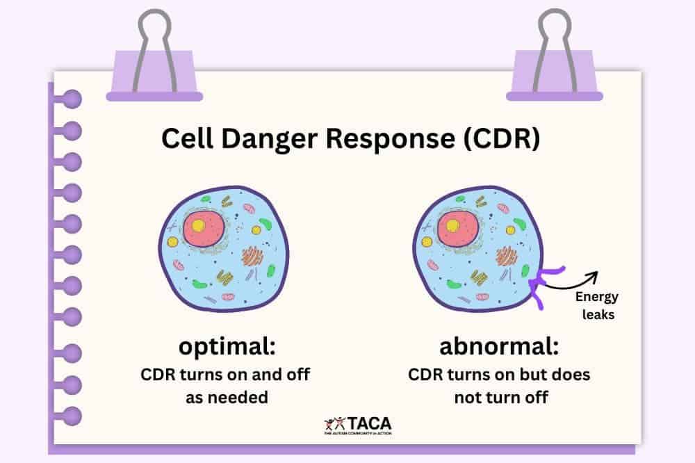
The CDR is the body’s ancient, conserved response to threat. When cells detect danger, such as infection, toxin, or injury, they shift into a defensive metabolic state. Energy production is reduced, cellular communication slows down , and cells essentially go quiet to protect themselves.
This is adaptive in the short term. After an acute COVID infection, you want this response. It’s doing its job.
The problem is when the CDR doesn’t turn off. When the body’s threat detection system remains activated long after the initial danger has passed, you get persistent metabolic suppression. This manifests as fatigue that doesn’t resolve with rest, cognitive dysfunction that doesn’t respond to sleep, and a nervous system that stays in a low-grade defensive posture.
Most Long COVID clinicians are not testing for or thinking about CDR biology. Many deal with chronic inflammation and persistent spike protein, but never take that next step to address chronic CDR.
2. They Don’t Understand Senescent Cells
Cellular senescence, what some researchers call the “zombie cell” problem, is another mechanism that rarely appears in standard Long COVID treatment frameworks, although it may accidentally be addressed (albeit not thoroughly enough) through treatments with other prescribed purposes.
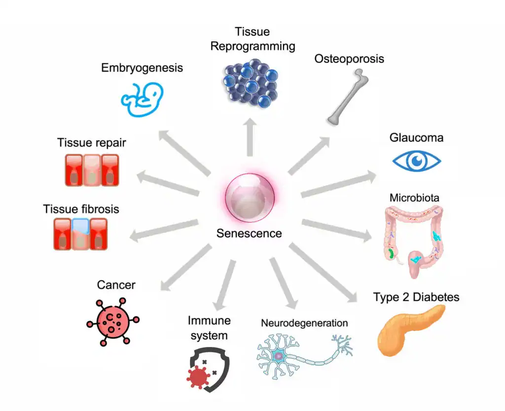
Senescent cells are cells that have stopped dividing but refuse to die. In the aftermath of a severe immune activation like COVID infection, the body can accumulate these cells in significant numbers. In fact, the spike protein seems to have be very skilled at producing senescent cells. These cells don’t do their normal jobs, but they also release a continuous stream of pro-inflammatory signaling molecules, the senescence-associated secretory phenotype (or SASP).
The result of senescent cell accumulation is a low-grade, chronic inflammatory state that can persist for years. Standard anti-inflammatory approaches don’t clear senescent cells. Senolytics, which are treatments that induce apoptosis in senescent cells, are rarely considered in clinical practice, let alone in Long COVID or Post-Vaccine Syndrome.
For patients where senescent cell burden is a significant contributor, which is all spike protein patients, treating everything else while ignoring this mechanism is like bailing water from a boat without fixing the leak.
3. They Apply Uniform Protocols to Non-Uniform Patients
We understand why this happens. Protocols are efficient. They can be standardized, taught, and scaled. If you’re running a practice that sees 50 Long COVID patients a week, a decision tree makes sense. Our practice model focuses on lower patient volumes for more personalized care and treatment. Why?
Long COVID patients are not uniform. The neurotransmitter profile of one patient – the specific pattern of catecholamine deficiency, glutamate dysregulation, and kynurenine pathway disruption – may look nothing like the next patient’s. The autoimmune burden, the degree of microclotting, the level of residual spike protein activity, the autonomic dysfunction pattern, may all vary enormously from patient to patient.
Our practice partner Scott Marsland, FNP-C, published a detailed case study that illustrates this complexity in just one patient, examining neurotransmitter changes across time and in response to specific interventions. The data showed 13 of 27 measured neurotransmitters were outside optimal range at baseline. The specific pattern of elevated glutamate suggesting excitotoxicity, paradoxically elevated serotonin alongside low tryptophan, and depleted catecholamines required a tailored response, not a standard protocol. And the picture changed meaningfully over seven months, requiring ongoing adjustment.
That is what individualized Long COVID care actually looks like.
4. They Over-Test and Under-Treat
There are particular kind of specialty practices that run $2,000–$5,000 in laboratory panels before doing much of anything. The testing is framed as “comprehensive” and “data-driven.” It feels thorough. As humans, we want to latch onto something concrete, like lab results. It gives us something to work at, and improve upon.
But here’s the honest clinical reality: in most Long COVID and PACVS patients, extensive testing rarely changes the initial treatment approach. The patterns we see across 3,500+ cases are recognizable. The likely mechanisms become apparent through careful history, symptom characterization, and clinical pattern recognition long before any lab results return.
Excessive testing delays treatment. It costs patients money they could have spent on interventions. And it creates an illusion of certainty in a condition that requires the intellectual honesty to acknowledge: we are treating this by thoughtful trial, observation, and adjustment. Running a panel and following a chart does not work for complex conditions like Long Covid and Post-Vaccine Syndrome. One day might turn up a normal lab result, and then many abnormalities the next testing period. Patients end up chasing ghosts.
And, by the way, if and when a $4,000 testing panel arrives that meaningfully guides pin-pointed treatment decisions tha deliver results, we will be the first ones to come back and edit this post.
5. They Treat the First Layer and Stop
Long COVID recovery, when it happens, typically unfolds across 9 to 18 months. Sometimes longer. The condition does not resolve in a single treatment arc, but requires ongoing attention to how the patient’s picture is shifting.
A treatment that was appropriate in month two may need modification by month five. An intervention that wasn’t relevant initially may become important as other issues resolve and different mechanisms become more apparent. The clinical work is iterative.
Many practices, particularly those operating at high volume or on a brief-consultation model, don’t have the infrastructure or clinical philosophy to support this. They prescribe an initial regimen and check in only when you schedule your next appointment. That interval is too long, and the flexibility to pivot is often not there.
What a Different Approach Looks Like
We want to be direct about what we actually do differently, not just what we criticize.
We start with clinical reasoning, not a panel. The first consultation at Leading Edge Clinic is 60 minutes. We are building a detailed picture of your symptom history, your illness trajectory, what you’ve tried, how you responded, and what the pattern suggests. That reasoning guides our initial approach, not a lab panel.
We think about mechanisms, not just symptoms. If you have fatigue, we want to understand whether it’s primarily metabolic, autonomic, inflammatory, or driven by a cellular danger response that hasn’t resolved. Different mechanisms call for different interventions.
We are honest about the trial-and-error nature of treatment. We don’t tell patients we’ve found the answer and here it is. We tell them: here is our best clinical hypothesis, here is the treatment we think is most likely to move things in the right direction, and here is how we’ll know if it’s working. We adjust based on what we observe.
We treat the evolving patient, not the initial presentation. Follow-up is built into how we work. As patients respond, we incorporate that information and adapt.
We consider mechanisms that others don’t. Cell danger response, cellular senescence, persistent spike protein activity, microclotting, neurological changes, autonomic dysfunction, immune dysregulation, MCAS, and other spike protein pathologies are all part of our clinical thinking. Not for every patient, but for the patients where these mechanisms are relevant, addressing them can be the difference between continued decline and meaningful recovery.
What You Should Ask Any Long COVID Specialist
If you’re evaluating whether a Long COVID practice is right for you, here are honest questions worth asking:
“What do you think is driving my specific symptoms?” A clinician who can give you a mechanistic hypothesis, specific to your history, and not a generic answer, is thinking carefully. A clinician who gives you the same answer they give everyone is running a protocol.
“What do you do when a treatment isn’t working?” The answer should involve active pivoting, consideration of alternative mechanisms, and willingness to try something different.
“How do you think about conditions like cell danger response or cellular senescence?” You’re not expecting a dissertation. But a blank look or a dismissive response tells you something about the depth of the clinical framework.
“How long do you expect treatment to take?” Honesty here matters. Anyone promising significant recovery in 6 to 8 weeks is either treating very mild cases or not being straight with you. The realistic timeline for meaningful improvement in complex Long COVID is 9 to 18 months.
A Note on What We Don’t Promise
We’ve seen enough patients and enough trajectories to say this plainly: approximately 80–85% of our patients achieve significant functional improvement over the course of treatment. That is a meaningful number, and we’re proud of it.
But 80–85% also means that 15–20% of patients don’t reach that threshold, at least not within our treatment window.
What we can promise is clinical honesty, genuine intellectual engagement with your case, and a willingness to keep thinking when the obvious approaches aren’t working.
If You’ve Already Tried Everything
If you’ve been through the conventional system, the university Long COVID clinics, the integrative medicine practices, the telehealth protocols – and you’re still significantly impaired – there may be value in a clinical framework that explicitly accounts for the mechanisms most others aren’t addressing.
We recognize that many of our patients come to us precisely because they’ve exhausted the obvious options and need something different.
A 60-minute initial evaluation gives us the opportunity to build a complete picture of your case: what you’ve tried, how you responded, what your symptom pattern suggests, and where we think there may be unexplored mechanistic territory. From that, we develop a realistic treatment roadmap, not a protocol applied to a category, but a clinical plan developed for your specific situation.
Long COVID and Post-Vaccine Syndrome not one disease. They don’t have one mechanism, they don’t respond to one protocol, and they don’t resolve on a predictable timeline.
The clinicians who are having the best outcomes with this condition are the ones who understand complexity, think in terms of mechanisms, are willing to adapt, and have the clinical experience to recognize patterns that don’t fit neatly into any category.
That is the medicine we practice. If you’ve been failed by other approaches and you’re still looking for meaningful progress, we’d be glad to talk.
Leading Edge Clinic specializes in Long COVID, Post-Vaccine Syndrome, and complex post-infectious illness. Our clinic has treated more than 3,500 patients with these conditions.
This article is for informational purposes and does not constitute medical advice. Individual results vary.
We have treated over 3,500 Long COVID and Post-Vaccine Syndrome cases since 2022. However, still, the treatments showing the most promise in clinical practice aren’t yet in the official guidelines.
This isn’t surprising. The gap between clinical innovation and institutional approval often takes years, sometimes decades, to close. That is, if they ever close at all. There is, of course, story after story of promising curative treatments purchased by pharmaceutical companies and immediately thrown in the filing cabinet of oblivion – innovation killed and stifled to maintain cash cow products that produce no meaningful results.
Now infamous, the time a Goldman Sachs analyst asked the quiet part out loud: “Is curing patients a sustainable business model?” The analyst when on to write in her report, “…such (curative) treatments offer a very different outlook with regard to recurring revenue versus chronic therapies. While this proposition carries tremendous value for patients and society, it could represent a challenge for genome medicine developers looking for sustained cash flow.”
Long COVID and Post-Vaccine Syndrome patients can’t wait to see if someone looks favorably at the economic viability of helping them with their severe and debilitating conditions. And if that endeavor is decidedly profitable (which it most certainly will be based on the current trajectory of severely ill spike protein patients), they can ill afford to wait the additional 5+ years for clinical research and testing for expensive, patented drugs that treat symptoms.
What follows is an evidence-based discussion of a small handful of treatments we’ve used successfully in clinical practice, why they work mechanistically, what the research shows, and why most physicians haven’t adopted them yet. This is by no means an exhaustive list. But, some treatments we thought were worth highlighting for one reason or another.
This represents real-world clinical experience combined with emerging research. The kind of information you need to have informed conversations with your healthcare provider.
Understanding the Treatment Gap
Before discussing specific treatments, it’s important to understand why there’s often a delay between what helps patients and what becomes “standard of care.”
Three Primary Barriers:
1. Regulatory Framework Most of these treatments are FDA-approved for other conditions but used “off-label” for Long COVID. While legal and common in medicine, off-label use requires physician comfort with clinical decision-making beyond established protocols. We saw during the pandemic that “off-label” suddenly became a dirty word, weaponized against certain drugs demonstrating promise in treatment. More on that later.
2. Evidence Timeline Rigorous clinical trials take 3-5 years from design to publication. Long COVID emerged in 2020. We’re only now seeing results from the first wave of controlled trials. Furthermore, these trials are often on patented therapies with high side effect profiles and little clinical efficacy.
3. Risk-Benefit Calculation Physicians must weigh potential benefit against professional liability, especially for treatments lacking specific FDA approval for Long COVID. Even when clinical rationale is strong. To us, the decision is easy. Integrity trumps all risk.
The result: Patients often wait years for treatments that clinical experience suggests could help now.
Treatment #1: Low-Dose Naltrexone (LDN)
What it is: Naltrexone at standard doses (50-100mg/day) treats opioid addiction. At lower doses (1-4.5mg/day), it functions as an immunomodulator with anti-inflammatory properties.
Clinical rationale for Long COVID:
Low-dose naltrexone works through multiple mechanisms relevant to Long COVID pathophysiology:
Immune modulation: Inhibits pro-inflammatory toll-like receptor 4 (TLR4), which drives cytokine production
Cytokine reduction: Decreases inflammatory markers including IL-6, TNF-alpha, and IL-1β
Ion channel restoration: A 2025 study demonstrated LDN restored TRPM3 ion channel function in Long COVID patients’ immune cells—significant because TRPM3 dysfunction impairs cellular energy production and immune response
Mast Cell Stabilization: By reducing immune reactivity and cytokine production, it also creates a more stabile environment, preventing histamine release from mast cells.
Neuroinflammatory Reduction: Reduces microglial activation in the brain, with 60% of users reporting reduction in anxiety, fatigue, and brain fog
Current evidence:
The NIH RECOVER program selected LDN for clinical trials, with enrollment beginning summer 2026. Existing data includes:
Meta-analysis of observational studies (n=155 across 4 studies): Moderate effect size for fatigue reduction (Hedges’ g = -0.74, p<0.001)
Quality of life improvements: SF-36 scores increased from 36.5 to 52.1 (p<0.0001) over 12 weeks
TRPM3 restoration study (Frontiers in Molecular Biosciences, 2025): Demonstrated objective improvement in immune cell calcium signaling
Safety profile: Excellent across multiple studies. Most common side effects are mild and transient (vivid dreams, slight headache in first 1-2 weeks)
Contraindications: Active opioid use, liver dysfunction
Clinical experience: In our practice, approximately 50-60% of Long COVID and Post-Vaccine Syndrome patients experience significant benefit from LDN. Response is most notable for fatigue, post-exertional malaise, and brain fog.
Typical response timeline: 8-12 weeks at therapeutic dose.
Prescribing approach:
Start low (0.5-1mg) to assess tolerance
Titrate slowly: increase by 0.5mg every 3-5 days
Target dose: 3-4.5mg daily
Taken at bedtime (may initially cause vivid dreams)
Requires compounding pharmacy
Why adoption is limited: Requires off-label prescribing, compounding pharmacy access, and physician comfort with immunomodulation. Many physicians prefer to wait for RECOVER trial results before prescribing. To us, it is shocking that it has taken this long to even get studies going on this low risk, high benefit option.
Treatment #2: Low-Dose Ketamine
What it is: Ketamine is an NMDA receptor antagonist originally used for anesthesia, now FDA-approved for treatment-resistant depression via the esketamine nasal spray (Spravato). We have written more extensively about its history, its mechanisms of action, and what we’ve seen in our Long Covid and Post-Vaccine Syndrome patients here
Clinical rationale for Long COVID and Post-Vaccine Syndrome:
Ketamine addresses multiple Long COVID and PACVS mechanisms:
Neuroinflammation reduction: Downregulates inflammatory cytokines (IL-6, IL-17A, TNF-alpha) that contribute to brain fog and cognitive dysfunction
Rapid-acting antidepressant: Unlike traditional SSRIs requiring 4-6 weeks, ketamine can show effects within hours to days, and takes a fundamentally different approach from serotonin based treatments
Chronic pain modulation: Effective for neuropathic pain through NMDA receptor antagonism
Current evidence:
Active clinical trials: University of British Columbia Phase 2 trial testing ketamine for Long COVID fatigue and cognitive symptoms (NCT identifier pending, 20 participants)
Case reports: Published cases demonstrating rapid improvement in treatment-resistant depression and suicidality in Long COVID patients. These reports are focused on psychological impacts. However, there are significant physiological benefits to treatment as well
Mechanism studies: Ketamine reduces specific inflammatory markers that correlate with Long COVID symptom severity
Combination therapy: German observational study showed promising results combining ketamine with repetitive transcranial magnetic stimulation (rTMS)
Administration routes:
Low-Dose Sublingual Ketamine: Although there are other administrative routes (IV, intranasal, etc…), to achieve therapeutic dosing for Long Covid and Post-Vaccine Syndrome patients, only low doses are needed. These doses can be achieved through sublingual administrations via compounded drops, or troches.
Clinical experience: Most dramatic improvements occur in patients with:
Severe fatigue with cognitive dysfunction
Chronic neuropathic pain
Persistent brain fog unresponsive to other interventions
Anxiety and depressive related disorders
Dysautonomia symptoms
Typical protocol: Daily sublingual drops or troches at minimally tolerable dose. Dosing dependent upon tolerability is based on glutamate imbalances, which are corrected over time with sublingual low-dose ketamine. Improvements are seen over a 6 month period.
Safety considerations:
Requires medical supervision and monitoring
Potential side effects with low-dose sublingual ketamine: Dissociation, minor euphoric feeling, anxiety during infusion
Contraindications: Active substance abuse (however, this can also be a treatment for substance abuse)
Not appropriate for all patients
Why adoption is limited: Off-label prescribing of low-dose sublingual ketamine for Long Covid and Post-Vaccine Syndrome is not something many clinicians are aware, let alone comfortable with. Its benefits are well-known in some psychiatric medicine circles for treatment resistant depression, but its effects on BDNF and neuronal healing are not yet mainstream for the treatment of other conditions, such as Long Covid, that impact brain health. It is a controlled substance, which means prescribers must have DEA licenses.
Treatment #3: Dimethyl Sulfoxide (DMSO)
What it is:Dimethyl sulfoxide(DMSO) is a chemical solvent and prescription medication primarily used to treat painful bladder syndrome (interstitial cystitis). It is known for its ability to rapidly penetrate skin and membranes, acting as an anti-inflammatory and analgesic agent.
Clinical rationale for Long COVID and Post-Vaccine Syndrome:
Some argue that DMSO is the most suppressed therapeutic in modern medicine because it works too well for too many things. You’ll remember from earlier in this article what the Goldman Sachs analyst said about effective, curative treatments. When the FDA banned DMSO in 1965 (despite overwhelming safety data), thousands of patients with debilitating conditions lost access to a therapy that was giving them their lives back. The parallels to how Long COVID and Post-Vaccine Syndrome patients are being dismissed today are not coincidental.
Here’s what makes DMSO uniquely suited for spike protein disease:
The Spike-Protein Connection: Why DMSO Makes Mechanistic Sense
If persistent spike protein is driving Long COVID and Post-Vaccine Syndrome pathology—and mounting evidence suggests it is—then DMSO addresses nearly every downstream mechanism we see clinically:
1. Protein Misfolding and Amyloid Fibrin Microclotting
DMSO is a chemical chaperone. It stabilizes protein folding and, critically, dissolves amyloid aggregates.
At least 40 studies have demonstrated DMSO’s ability to solubilize amyloid fibrils and enable the body to break them down. If spike protein is creating misfolded proteins or amyloid-like microclots (the leading hypothesis for many Long COVID and PACVS symptoms), DMSO has a direct mechanism of action:
Prevents proteins from aggregating into pathologic forms
Dissolves existing amyloid deposits
Allows the body to clear these aggregates through normal elimination pathways
This isn’t theoretical, or even in vitro evidence. DMSO has successfully treated amyloidosis in humans—a condition characterized by insoluble protein aggregation causing organ damage. Same mechanism, different protein.
The microclotting angle: DMSO is also a powerful platelet deaggregator and anticoagulant:
Inhibits platelet aggregation induced by ADP, collagen, arachidonic acid, and platelet-activating factor
Blocks tissue factor (TF) expression—the key link between inflammation and clotting
Unlike aspirin or heparin, it does this without bleeding risk
If microclots are clogging your microcirculation and causing tissue hypoxia, DMSO addresses it directly while simultaneously improving blood flow to oxygen-starved tissues.
2. Autoimmunity
Spike-triggered autoimmunity is one of the most devastating Long COVID mechanisms. DMSO has decades of evidence treating autoimmune conditions that conventional medicine still can’t touch:
Lupus – reduces symptoms, often allowing steroid reduction
Multiple sclerosis – stops progression in many cases, improves symptoms
Rheumatoid arthritis – 70-80% response rate in multiple studies
Scleroderma – one of the few things that works for this “untreatable” condition
Interstitial cystitis – the only FDA-approved use for DMSO (because the evidence was impossible to suppress)
Sjögren’s syndrome – dramatically improves dry mouth/eyes in published case series
Improves microcirculation – documented in studies using radioactive isotope imaging
Treats vasospasm – successfully used for Raynaud’s syndrome, peripheral neuropathy, complex regional pain syndrome
Clinical studies show DMSO:
Eliminates symptoms in 50% of Raynaud’s patients
Treats diabetic peripheral neuropathy and prevents amputations
Resolves varicose veins (sometimes within minutes of IV administration)
Improves circulation in thrombophlebitis and peripheral artery disease
If spike-ACE2 interaction is damaging your vasculature, DMSO protects and heals blood vessels.
4. Neurological Protection and Small Fiber Neuropathy
Small fiber neuropathy is the #4 most common vaccine injury symptom. It’s also prevalent in Long COVID.
DMSO selectively blocks C-fibers and A-delta fibers—the exact nerve fibers responsible for burning pain, electrical shocks, pins-and-needles, and the agonizing dysesthesias of small fiber neuropathy.
Mechanism:
Blocks nerve conduction in small pain fibers without affecting larger motor/sensory fibers
Suppresses NMDA and AMPA receptor activity (central pain sensitization)
Blocks excessive calcium/sodium influx into pain-transmitting neurons
Does NOT create tolerance (unlike opioids—often becomes MORE effective over time)
Additionally, DMSO crosses the blood-brain barrier and:
Protects neurons from death following ischemia/hypoxia
Reduces neuroinflammation
Treats brain fog, cognitive dysfunction (reported extensively in Long COVID patients using DMSO)
Has successfully treated traumatic brain injuries, strokes, spinal cord injuries in studies the FDA ignored
5. Organ Protection and Healing
DMSO doesn’t just reduce symptoms, it heals damaged tissue. This is critical for Long COVID and Post-Vaccine Syndrome patients with organ involvement (which would be most patients):
Lungs:
Reduces pulmonary fibrosis (case reports of transplant candidates recovering)
Treats COPD, asthma exacerbations
Improves oxygenation in respiratory insufficiency
81% of patients with chronic respiratory failure improved without hospitalization in one study
Dosing: We start with topical and/or oral dosing based on specific patient needs, titrating appropriately based on response and tolerability. Some patients seek out IV administration for more aggressive treatment, though this is logistically challenging outside specialized clinics.
Expected timeline:
Acute symptoms (pain, inflammation): Often improve within hours to days
Chronic conditions: 4-7 days to start seeing benefit, 6-8 weeks for sustained improvement
DMSO often becomes MORE effective with continued use (opposite of pharmaceutical tolerance)
Contraindications and cautions:
Pregnancy/breastfeeding – inadequate safety data (though one study showed safe use for infertility)
Active skin infections – DMSO can carry surface toxins into the body; ensure skin is clean before application
Severe liver or kidney dysfunction – use with caution, monitor closely
Concurrent DMSO + PRP injections – may reduce PRP efficacy due to platelet inhibition
Allergic reactions – rare (~1 in 2000) but possible; start with low concentration to assess tolerance
Skin irritation with topical use (concentration-dependent, often resolves with continued use)
Nausea (uncommon with appropriate dosing)
Why You Haven’t Heard About This
The FDA banned DMSO research in 1965 based on a fabricated safety concern (lens opacities in dogs—which never occurred in any other species or in humans). Despite Congressional hearings, outcry from patients and physicians, and a former Secretary of Health and Human Services championing it after using DMSO to treat his wife’s terminal cancer pain, the FDA never relented.
Why? DMSO threatened too many profitable drug markets.
In 1966, over 1,000 researchers presented evidence at the Waldorf Astoria. Zero withdrew their papers despite FDA pressure. The data was overwhelming.
Sixty years later, patients are still suffering from conditions DMSO could treat—because the FDA decided your access to effective medicine was less important than protecting pharmaceutical profits.
For Long COVID patients failed by the medical establishment, DMSO represents what medicine should have been: safe, effective, accessible, and focused on healing rather than management.
Bottom line on DMSO for Long COVID:
If persistent spike protein is causing:
Protein misfolding → DMSO refolds and stabilizes proteins
Amyloid microclots → DMSO dissolves aggregates and prevents platelet aggregation
Organ damage → DMSO protects lungs, gut, heart, kidneys
The mechanism isn’t speculative. The evidence isn’t anecdotal. This is decades of suppressed research finally reaching patients who need it.
Treatment #4: Microcurrent Therapy
What it is: Microcurrent therapy delivers sub-sensory electrical currents in the microampere range — currents so small they mimic the body’s own bioelectrical signals. We commonly have recommended the Arc Microtech device. The Arc device was engineered by Ian Thirkell, a retired English detective who spent years studying the bioelectricity research of Dr. Robert O. Becker after his wife handed him three of Becker’s books and told him to go learn something useful.
The scientific foundation is worth understanding. Becker was an orthopedic surgeon and researcher at the VA Hospital in Syracuse, New York, who spent decades studying how the body uses electrical signals to heal. He discovered that injury generates a measurable shift in voltage at the wound site — a “current of injury” — that initiates the healing process. He demonstrated that applying tiny electrical currents could stimulate partial limb regeneration in rats (published in Nature, 1972) and developed techniques using electrically generated silver ions to fight antibiotic-resistant infections and promote tissue regeneration in human patients. His core insight, detailed in his landmark book The Body Electric (1985): the body operates on a bioelectrical control system that can be supported and restored through the careful application of currents matching the body’s own frequencies. The ARC device is built directly on this principle.
Clinical rationale for Long COVID and Post-Vaccine Syndrome:
ATP production enhancement: The landmark 1982 study by Cheng et al. demonstrated that microcurrent stimulation in the 100-500 microampere range increased ATP production by up to 500% in treated tissue, while increasing amino acid transport by 30-40% and enhancing protein synthesis. When current was increased into the milliampere range used by TENS units, ATP production actually decreased. This distinction matters enormously: Long COVID and Post-Vaccine Syndrome patients have documented mitochondrial dysfunction. A 2025 study in Annals of Medicine confirmed that immune cells from Long COVID patients exhibit aberrant ATP synthase function, resulting in diminished cellular energy availability. The spike protein — whether from viral reservoirs or circulating post-vaccination — impairs oxidative phosphorylation, the process responsible for 90% of cellular energy production. Fatigue, post-exertional malaise, brain fog, exercise intolerance — these are manifestations of cells that cannot produce adequate energy (although there is greater detail as to what is happening here, some of this is detailed in our prior article on cell danger response). Microcurrent directly targets this deficit.
Inflammation reduction: Microcurrent reduces inflammatory markers and promotes resolution of swelling through enhanced circulation and lymphatic drainage — relevant to the chronic systemic inflammation driving Long COVID and Post-Vaccine Syndrome symptoms
Tissue repair and cellular regeneration: Building on Becker’s work, microcurrent stimulates fibroblast activity, enhances collagen synthesis, and promotes angiogenesis. For patients with endothelial damage and microclotting from spike protein, these repair mechanisms are critical. We have seen patients in heart failure restore their ejection fraction back to well over the CHF baseline.
Autonomic and neurological support: Clinical observations from our practice and colleagues in the UK suggest meaningful benefit for patients with autonomic dysfunction, anxiety, depression, and PTSD — conditions that frequently accompany Long COVID and Post-Vaccine Syndrome
One framing we find useful: supplements that support mitochondrial function — CoQ10, B vitamins, magnesium — are static interventions for what is fundamentally a dynamic process. They provide raw materials. Microcurrent is a dynamic intervention: it actively stimulates the cellular machinery to do its job. The two approaches are complementary, not competing.
Current evidence:
Cheng et al. (1982): Foundational study demonstrating 500% ATP increase at therapeutic microcurrent levels, with concurrent increases in amino acid transport and protein synthesis Becker’s published work (1961-1998): Decades of peer-reviewed research on bioelectrical control of regeneration, published in Nature, Science, and other major journals Modern reviews: A 2025 narrative review confirmed microcurrent’s role in enhancing ATP synthesis, improving mitochondrial efficiency, and activating tissue repair mechanisms No randomized controlled trials specific to microcurrent for Long COVID exist, and they may never — putting it in the same category as most treatments on this list when they were first adopted clinically.
Clinical experience: In our practice, over 500 patients have used the ARC device. It has been a reliable and safe recommended interventions for a few years now.
Most consistent improvements:
Fatigue — the most reliably reported benefit, typically noticeable after approximately six weeks of consistent use Pain and inflammation — reduced swelling and bruising, notably effective for patients on anticoagulation therapy Cardiac function — we have observed cases of improved cardiac ejection fraction with sustained use, corroborated by colleagues in the UK Brain fog and cognitive function — improvements likely related to enhanced cellular energy production and reduced neuroinflammation Anxiety and trauma-related symptoms — consistent with observations from military psychiatrists using microcurrent for treatment-resistant PTSD
Benefit is cumulative. More consistent use produces greater results.
Typical protocol:
Program 1 (100% anti-inflammatory): At least one 3-hour cycle daily for two months Program 2 (50% anti-inflammatory, 50% cellular repair): At least one 3-hour cycle daily for two weeks Program 3 (25% anti-inflammatory, 75% cellular repair): At least one 3-hour cycle daily for two weeks Then rotate: One week each of Programs 1, 2, and 3, repeating continuously
Worn on the arm or leg (most patients prefer above the ankle). The strap should be fitted but not tight — two fingers should fit between strap and skin. Tightness inhibits circulation and reduces benefit.
Response timeline: Most patients notice benefit after approximately six weeks. Some report improvement within days, particularly for pain and inflammation. However, some patients even report improved energy in just days.
Safety considerations:
Excellent safety profile — these are sub-sensory currents operating at the same magnitude as the body’s own cellular electrical signaling Approximately 5% of patients experience initial sensitivity (nausea, dizziness), managed by starting with very short sessions and gradually increasing No significant adverse effects observed in our patient population Safe for use alongside other treatments, including anticoagulation Contraindications: Active implanted electrical devices (pacemakers, defibrillators)
Why adoption is limited: Microcurrent therapy sits entirely outside the pharmaceutical paradigm. There is no drug to prescribe, no procedure to bill for, and no pharmaceutical company funding trials. Becker himself faced significant institutional resistance throughout his career — his research challenged the chemical-mechanistic model of biology, and his opposition to electromagnetic pollution put him at odds with powerful interests. The ARC Microtech is a small, family-owned UK company without resources for large-scale clinical trials. And the concept that a wearable device delivering imperceptible electrical currents can meaningfully impact chronic illness strikes most conventionally trained physicians as implausible, despite decades of published research supporting the underlying science.
Our perspective: Microcurrent therapy, and specifically the ARC device, is one of the most underappreciated tools in our clinical arsenal. It is non-invasive, has an excellent safety profile, produces cumulative benefit, and directly addresses what may be the single most important pathological mechanism in Long COVID and Post-Vaccine Syndrome: mitochondrial energy failure. For a condition where the body has lost its ability to produce adequate energy, providing it with the bioelectrical signal to restore that production isn’t alternative quackery — it’s the application of proven biophysics.
Conflicts of Interest: We do offer a discount code for the ARC Microtech device. Using code USARCLEC at checkout on the ARC website provides a $20 discount to ordering customers. ARC also provides us with $20. However, we have never stashed this money away in a bank account. We have historically used this money to re-invest into devices for patients who can not afford to invest in this special device.
Treatment #5: Ivermectin
The controversial treatment that requires honest discussion.
Ivermectin became one of the most politicized medications in modern history. That makes it difficult to have rational, evidence-based conversations about its potential role in Long COVID and Post-Vaccine Syndrome.
Here’s what we actually know:
What it is: An antiparasitic medication on the WHO’s List of Essential Medicines, with an excellent 40-year safety record from billions of doses administered globally.
Theoretical mechanisms for Long COVID and Post-Vaccine Syndrome:
1. Spike Protein and ACE2 Receptor Interaction: This is perhaps the most relevant mechanism for Long COVID and Post-Vaccine Syndrome. Multiple molecular docking studies have demonstrated that ivermectin binds to:
The spike protein receptor-binding domain (RBD): Where the spike protein attaches to ACE2 receptors
The spike-ACE2 complex interface: Specifically between leucine 91 of the spike protein and histidine 378 of the ACE2 receptor
The N-terminal domain (NTD): Which controls initial viral attachment to cell membranes
Binding energy studies show strong affinity (-18 kcal/mol), suggesting ivermectin may physically interfere with spike protein attachment to ACE2 receptors.
Why this matters for Long COVID and Post-Vaccine Syndrome: If persistent spike protein (either from viral reservoirs or circulating spike) is driving symptoms—a leading hypothesis in Long COVID and Post-Vaccine Syndrome pathophysiology—ivermectin’s ability to bind both spike protein and ACE2 receptors could theoretically:
Reduce spike-mediated inflammation and endothelial damage
2. Additional antiviral mechanisms:
TMPRSS2 binding: Ivermectin binds to TMPRSS2, a protease that activates spike protein for cell entry
Viral replication inhibition: Demonstrated in vitro inhibition of SARS-CoV-2 replication
Multiple viral protein targets: Binds to main protease (Mpro), RNA-dependent RNA polymerase (RdRp), and nucleocapsid proteins
3. Anti-inflammatory effects:
Inhibits nuclear import of inflammatory transcription factors
Reduces cytokine production (IL-6, TNF-alpha, IL-1β)
Modulates immune response through multiple pathways
Important concentration caveat: While docking studies predict strong binding, debate exists about whether therapeutically achievable concentrations in humans are sufficient for these effects. One 2024 study suggested effective spike binding required concentrations 100-1000x the approved dosage in their assay system, though other studies and clinical observations suggest benefit at standard doses. Our clinical observations suggest extremely safe dosing ranges are effective in about 70% of patients.
The discrepancy may relate to:
Differences between in vitro assays and in vivo conditions
Tissue concentration versus serum concentration
Individual pharmacokinetic variation
Duration of exposure in chronic dosing versus single-dose studies
The evidence landscape:
The data on ivermectin for acute COVID-19 was mixed, politicized, and contentious. For Long COVID specifically:
No published randomized controlled trials
Mechanistic rationale exists
Clinical experience varies among practitioners. Our experience suggests a 70% positive clinical response rate
Clinical experience: In our practice, we’ve observed a subset of patients—approximately 70%—who report improvement when ivermectin is included as part of a comprehensive treatment protocol.
Important caveats:
Not a monotherapy; used as part of multi-modal approach
Standard anti-parasitic dosing (not megadoses)
Drug interactions exist
Why adoption is limited: The political controversy surrounding ivermectin creates significant professional risk for physicians who prescribe it, even for legitimate clinical indications. Many state medical boards have issued warnings, and physicians face potential peer censure. Dr. Kory had his board certifications revoked for promoting its use in acutely ill COVID patients, even in spite of positive clinical responses.
Our perspective: With Dr. Kory having testified before the Senate about COVID treatments and faced professional consequences for advocating treatments outside the mainstream, we believe in the efficacy of ivermectin based on excellent safety profile, low cost, and high clinical response rates.
The question for any treatment should be: What does the evidence show, what is the safety profile, and what is the clinical experience?
For ivermectin in Long COVID and Post-Vaccine Syndrome: Mainsteam evidence is limited, the safety profile is excellent, and our clinical experience is positive.
Whether to try it should be an informed decision between patient and physician, weighing potential benefits against minimal risks.
Treatment #6: Anticoagulation Therapy
What it is: Blood-thinning medications ranging from aspirin to low-dose anticoagulants.
Clinical rationale:
Growing evidence suggests microclots may play a role in Long COVID:
Microclot hypothesis: Persistent microclotting causing tissue hypoxia and symptoms
Proven benefit in acute COVID: Anticoagulation improves outcomes in hospitalized patients
Current evidence: Mechanistically compelling. Clinical trials underway but results pending.
Clinical experience: Some patients show improvement with aspirin or low-dose anticoagulation. Requires careful patient selection and monitoring.
Safety considerations: Bleeding risk requires careful assessment. Not appropriate for all patients.
Why adoption is limited: Balancing benefit versus bleeding risk requires individualized assessment. Physicians appropriately cautious about anticoagulating without clear indication.
The Gap Between Evidence and Practice
Every treatment discussed here shares common characteristics:
Mechanistic rationale based on Long COVID and PACVS pathophysiology
Existing clinical experience supporting use
Acceptable safety profiles when used appropriately
Real clinical results within our treatment of over 3,500 Long Covid and Post-Vaccine Syndrome patients
Yet most physicians haven’t adopted them. Why?
The Institutional Reality:
Evidence requirements: Institutions typically require randomized controlled trials before treatment adoption. These trials take years.
Liability concerns: Off-label prescribing, while legal, increases perceived malpractice risk.
Guideline absence: Without official clinical guidelines, physicians lack institutional backing for treatment decisions.
Professional pressure: Medical boards, peer review, and institutional policies create incentives to wait for official recommendations.
This creates a gap: Patients suffering now while waiting for evidence that may take years to generate.
What You Should Know
These Treatments Aren’t Magic Bullets
Long COVID and Post-Vaccine Syndrome are heterogeneous and complex. Treatment response varies significantly between patients.
The ideal scenario would be completed randomized controlled trials for each treatment.
What we actually have:
Mechanistic understanding (how they should work)
Clinical experience (what we’ve observed)
Emerging trial data (what’s being formally tested)
Urgent patient need (can’t wait years for perfect evidence)
The challenge: Balancing the desire for perfect evidence with the reality of patients suffering now.
This Requires Specialized Care
Optimal Long COVID and Post-Vaccine Syndrome treatment requires a clinician who:
Understands complex post-viral illness
Can navigate off-label prescribing appropriately
Will monitor carefully for adverse effects
Knows when to adjust or discontinue treatment
Can integrate multiple therapeutic modalities
Not all physicians have this expertise or comfort level.
Clinical Outcomes
In our practice of 3,500+ Long COVID patients:
80-85% achieve significant functional improvement
This means:
Return to work (often with modifications initially)
Ability to exercise (frequently at reduced level initially)
Brain fog resolved or significantly improved
Fatigue reduced to manageable levels
Quality of life substantially better
Realistic timeline: 9-18 months on average
Long COVID and Post-Vaccine Sydnrome are chronic conditions requiring sustained treatment. Promises of rapid recovery are unrealistic for most patients.
Finding Appropriate Care
If you’re considering these treatments:
1. Informed discussion with your current physician Share this information. Ask if they’re willing to consider these approaches.
2. Seek Long COVID expertise Find physicians with specific experience treating post-viral illness.
3. Consider telemedicine options Some Long COVID and PACVS specialists work across state lines via telemedicine.
4. Join support communities Other patients can recommend physicians with Long COVID expertise.
5. Be your own advocate Come prepared with questions. Understand the rationale. Participate actively in treatment decisions.
The Bottom Line
Long COVID and PACVS treatment requires:
Integration of emerging research
Clinical judgment based on experience
Willingness to use approved medications for new indications
Patient-centered approach to risk-benefit analysis
Honest acknowledgment of uncertainty
Not all physicians are equipped or willing to practice this way. The traditional model of “wait for definitive evidence” serves patients poorly when that evidence is years away.
At our practice, we integrate the best available evidence with clinical experience from over 3,500 cases. We’re transparent about what we know, what we don’t know, and what we’ve observed.
This represents clinical medicine as it should be practiced: Thoughtful, evidence-informed, patient-centered, and unafraid of reasonable uncertainty.
If you’re a Long COVID or Post-Vaccine Syndrome patient seeking care that integrates clinical experience with emerging research, we treat complex post-viral cases at Leading Edge Clinic. Our approach is based on 3,500+ patient encounters, current medical literature, and individualized treatment planning.
Baldwin K, Wanson A, Gilecki L-A, Dalton C, Peters E, Halpape K. Intranasal ketamine as a treatment for psychiatric complications of long COVID: a case report. Mental Health Clinician. 2023;13(5):239-243. doi:10.9740/mhc.2023.10.239. PMC10732124. https://pmc.ncbi.nlm.nih.gov/articles/PMC10732124/
Evaluating the Neuromodulatory Effect of Ketamine in Long COVID-19. ClinicalTrials.gov Identifier: NCT06821087. University of British Columbia. https://clinicaltrials.gov/study/NCT06821087
Rolle C, Scheib M, Frank A, Russ I. Treatment of Chronic Fatigue Syndrome (CFS) in Post-SARS-CoV-2 Infection through combined outpatient Neuromodulation Therapy with Repetitive Transcranial Magnetic Stimulation (rTMS) and Ketamine IV Therapy — A Case Series. International Clinical Medical Case Reports Journal. https://ketaminplus.com/en/medical-studies
Zanos P, Moaddel R, Morris PJ, et al. Ketamine and ketamine metabolite pharmacology: insights into therapeutic mechanisms. Pharmacological Reviews. 2018;70(3):621-660. doi:10.1124/pr.117.015198.
Treatment #3: DMSO
Amyloid fibril dissolution / chemical chaperone:
Dzwolak W, Loksztejn A, Smirnovas V. Noncooperative dimethyl sulfoxide-induced dissection of insulin fibrils: toward soluble building blocks of amyloid. Biochemistry. 2009;48(26):6272-6284. https://pubmed.ncbi.nlm.nih.gov/19385641/
Kardos J, Yamamoto K, Hasegawa K, et al. Dissolution of beta2-microglobulin amyloid fibrils by dimethylsulfoxide. Journal of Biological Chemistry. 2003;278(24):21222-21227. https://pubmed.ncbi.nlm.nih.gov/12944383/
Hoshino M, Katou H, Hagihara Y, et al. Dimethylsulfoxide-quenched hydrogen/deuterium exchange method to study amyloid fibril structure. Biochimica et Biophysica Acta (BBA) – Biomembranes. 2007;1768(8):1886-1899. https://www.sciencedirect.com/science/article/pii/S0005273607000703
Iwai A, Yoshida T, Saito T, et al. Studies on biological actions of dimethyl sulfoxide in familial amyloidosis. Annals of the New York Academy of Sciences. 1983;411:52-64. https://pubmed.ncbi.nlm.nih.gov/6576722/
Amemori S, Iwakiri R, Ootani H, et al. Oral dimethyl sulfoxide for systemic amyloid A amyloidosis complication in chronic inflammatory disease: a retrospective patient chart review. Journal of Gastroenterology. 2006;41:444-449. https://link.springer.com/article/10.1007/s00535-006-1792-3
Asmis LM, Bazargan A, Pellegrin M, et al. DMSO inhibits human platelet activation through cyclooxygenase-1 inhibition. A novel agent for drug eluting stents? Biochemical and Biophysical Research Communications. 2010;391(4):1629-1633. https://pubmed.ncbi.nlm.nih.gov/20035720/
Saeed SA, Rasheed H, Ali TH, et al. Effects of dimethyl sulphoxide on aggregation of human blood platelets. Experimental and Molecular Pathology. 1987;46(2):159-169. https://pubmed.ncbi.nlm.nih.gov/2880990/
Rosenblum WI. Dimethyl sulfoxide effects on platelet aggregation and vascular reactivity in pial microcirculation. Annals of the New York Academy of Sciences. 1983;411:110-119. https://pubmed.ncbi.nlm.nih.gov/6410963/
Rosenblum WI. Dimethyl sulfoxide (DMSO) and glycerol, hydroxyl radical scavengers, impair platelet aggregation within and eliminate the accompanying vasodilation of, injured mouse pial arterioles. Stroke. 1982;13(1):35-39. https://www.ahajournals.org/doi/10.1161/01.str.13.1.35
Cheng N, Van Hoof H, Bockx E, et al. The effects of electric currents on ATP generation, protein synthesis, and membrane transport in rat skin. Clinical Orthopaedics and Related Research. 1982;(171):264-272.
Becker RO. Stimulation of partial limb regeneration in rats. Nature. 1972;235(5333):109-111.
Becker RO, Spadaro JA. Electrical stimulation of partial limb regeneration in mammals. Bulletin of the New York Academy of Medicine. 1972;48(4):627-641.
Becker RO, Chapin S, Sherry R. Regeneration of the ventricular myocardium in amphibians. Nature. 1974;248(444):145-147.
Becker RO, Selden G. The Body Electric: Electromagnetism and the Foundation of Life. William Morrow & Company; 1985.
Becker RO. Iontopheretic system for stimulation of tissue healing and regeneration. US Patent 5814094A. 1998.
Jonik S, Rothka AJ, Cherin N. Investigating the therapeutic efficacy of microcurrent therapy: a narrative review. Journal of Rehabilitation Medicine. 2025. PMC12357078. https://pmc.ncbi.nlm.nih.gov/articles/PMC12357078/
Piras A, Trofè A, Piperi I, et al. Physiological effects of microcurrent and its application for maximising acute responses and chronic adaptations to exercise. European Journal of Applied Physiology. 2022. https://link.springer.com/article/10.1007/s00421-022-05097-w
Lehrer S, Rheinstein PH. Ivermectin docks to the SARS-CoV-2 spike receptor-binding domain attached to ACE2. In Vivo. 2020;34(5):3023-3026. PMC7652439. https://pmc.ncbi.nlm.nih.gov/articles/PMC7652439/
Eweas AF, Alhossary AA, Abdel-Moneim AS. Molecular docking reveals ivermectin and remdesivir as potential repurposed drugs against SARS-CoV-2. Frontiers in Microbiology. 2021;11:592908. doi:10.3389/fmicb.2020.592908.
Ahmad S, Waheed Y, Abro A, Abbasi SW, Ismail S. Molecular screening of glycyrrhizin-based inhibitors against ACE2 host receptor of SARS-CoV-2. Structural Chemistry. 2021;32:1441-1452.
[2024 NTD binding study — Ivermectin binds spike N-terminal domain across variants including KP.3. Viruses (MDPI). 2024.]
Treatment #6: Anticoagulation Therapy
Kell DB, Laubscher GJ, Pretorius E. A central role for amyloid fibrin microclots in long COVID/PASC: origins and therapeutic implications. Biochemical Journal. 2022;479(4):537-559. doi:10.1042/BCJ20220016. https://portlandpress.com/biochemj/article/479/4/537/230829/
Pretorius E, Venter C, Laubscher GJ, et al. Prevalence of symptoms, comorbidities, fibrin amyloid microclots and platelet pathology in individuals with Long COVID/Post-Acute Sequelae of COVID-19 (PASC). Cardiovascular Diabetology. 2022;21(1):148. doi:10.1186/s12933-022-01579-5. https://link.springer.com/article/10.1186/s12933-022-01579-5
Pretorius E, Vlok M, Venter C, et al. Persistent clotting protein pathology in Long COVID/Post-Acute Sequelae of COVID-19 (PASC) is accompanied by increased levels of antiplasmin. Cardiovascular Diabetology. 2021;20:172. doi:10.1186/s12933-021-01359-7.
Grobbelaar LM, Venter C, Vlok M, et al. SARS-CoV-2 spike protein S1 induces fibrin(ogen) resistant to fibrinolysis: implications for microclot formation in COVID-19. Bioscience Reports. 2021;41(8):BSR20210611. doi:10.1042/BSR20210611.
Kruger A, Vlok M, Turner S, et al. Proteomics of fibrin amyloid microclots in long COVID/post-acute sequelae of COVID-19 (PASC) shows many entrapped pro-inflammatory molecules that may also contribute to a failed fibrinolytic system. Cardiovascular Diabetology. 2022;21(1):190. doi:10.1186/s12933-022-01623-4. PMC9491257. https://pmc.ncbi.nlm.nih.gov/articles/PMC9491257/
Libby P, Lüscher T. COVID-19 is, in the end, an endothelial disease. European Heart Journal. 2020;41(32):3038-3044. doi:10.1093/eurheartj/ehaa623.
Medical Disclaimer: This content is for educational purposes only and does not constitute medical advice. All treatments discussed require physician supervision. Consult a qualified healthcare provider before starting any new treatment. Individual results vary. Treatment decisions should be made in consultation with your physician based on your specific medical history and circumstances.
If you developed POTS (Postural Orthostatic Tachycardia Syndrome) after COVID infection or vaccination, you’ve probably been told to:
Drink more water and increase salt
Wear compression stockings
Try beta-blockers or midodrine
Do gradual exercise reconditioning
And maybe you’ve tried all of these. Some helped a little. But you’re still dealing with:
Racing heart when you stand up
Dizziness and lightheadedness
Extreme fatigue after being upright
Brain fog that worsens throughout the day
Feeling like you’re going to pass out
Significant symptom burden first thing in the morning
Here’s what we’ve learned after treating thousands of Long COVID and Post-Vaccine Syndrome patients with POTS:
Standard POTS treatments address the symptoms. But in 70% or more of the patients who come to us, there’s an underlying structural problem that’s driving the whole thing. A problem that most doctors don’t know to check for.
And when we fix this underlying problem? Many patients see dramatic improvement, sometimes within days of treatment.
The Hidden Problem Most Doctors Miss
After treating patients with post-viral and post-vaccine POTS since 2020, we’ve found something striking:
Over 70% of our POTS patients have significant venous compression in their pelvis.
Specifically, compression of the left common iliac vein, which is the major vein that drains blood from your left leg back to your heart.
This isn’t some fringe condition. Research shows:
69% of POTS patients have significant iliac vein compression (compared to 40% of healthy controls)
Up to 80% of POTS patients have some form of pelvic venous insufficiency
Yet most doctors never evaluate for it
Why does this matter?
Because if your POTS is being driven by poor venous return to your heart, all the salt and compression stockings in the world won’t fix the underlying problem.
It’s like trying to fill a bucket that has a hole in the bottom. You can keep pouring water (blood volume expansion, medications), but if the drainage system is blocked, you’re not solving the actual issue.
What Is Iliac Venous Compression?
Your left common iliac vein sits behind your right common iliac artery. In the normal anatomy, this is fine. Blood flows through both vessels without issue.
But in many people, especially after spike protein exposure from COVID or vaccination, the vein gets compressed by the artery sitting on top of it.
This compression is called May-Thurner Syndrome or iliac venous compression syndrome.
What happens when this vein is compressed?
Blood from your left leg can’t return to your heart efficiently
Blood pools in your lower body, worsened by postural changes such as standing
Less blood returns to your heart
Your heart has to beat faster to maintain blood pressure (hello, POTS)
You get all the classic POTS symptoms
Here’s the key insight: While this is an anatomical quirk that some people are born with, there still needs to be an activating event.
In Long COVID and Post-Vaccine Syndrome patients, spike protein is actively causing this problem through two main pathologies that impact the structural integrity of blood vessel walls.
— Clinical Observation from Leading Edge Clinic:
In our practice, we’ve observed that many Long COVID and Post-Vaccine Syndrome patients present with venous compression that wouldn’t have been clinically significant before spike protein exposure. The spike-induced endothelial and collagen dysfunction appears to transform anatomical variations that were previously asymptomatic into symptomatic compression syndromes.
One representative case: A 34-year-old female developed severe POTS 6 months post-vaccination. Imaging revealed 60% left common iliac vein compression—a finding that may have been present anatomically for years but only became symptomatic after spike protein triggered endothelial dysfunction and reduced her venous compliance.
Another case: A 30 year old male had a gradual onset of POTS that eventually became severe post-vaccination. Prior to Post-Vaccine Syndrome, patient was active, working out 6 days a week (hot yoga, kettlebell HIIT workouts, 30 mile cycling rides, etc…). A 3 day treatment of stem-cell exosomes improved some chest pain symptoms, but the POTS symptoms remained. Patient found mornings to be particularly difficult, with leg pain upon waking. Minor exertion caused continued chest pain and poor recovery. MRA and MRV revealed a significant stenosis indictative of IVC, which was confirmed by Dr. Brooke Spencer during an IVUS at stenting procedure. The IVUS showed an 80+% stenosis. POTS symptoms resolved upon stenting.
From the clinical observations of Dr. Scott Marsland, practice partner at Leading Edge Clinic —
The Spike Protein Connection
Why do so many Long COVID and Post-Vaccine Syndrome patients develop venous compression?
Two mechanisms:
1. Endothelial Dysfunction
Spike protein damages the endothelium (the inner lining of blood vessels), especially the glycocalyx. This causes:
Inflammation of vessel walls
Reduced elasticity
Impaired vascular function
Makes veins more susceptible to compression
Your veins rely on healthy endothelium to maintain their structure and resist external pressure. When spike protein damages this lining, the veins become weaker and more easily compressed.
2. Collagen Dysfunction
Spike protein also interferes with normal collagen synthesis and function. This creates a situation similar to what we see in Ehlers-Danlos Syndrome (EDS):
Weakened connective tissue
Reduced structural support for veins
Increased vein distensibility (they stretch and collapse more easily)
Higher risk of venous compression syndromes
Think about it like this: If the walls of your vein are weakened by spike-induced collagen dysfunction, they’re more likely to collapse under the pressure of the artery sitting on top.
This is why we see such high rates of venous compression in Long COVID and Post-Vaccine Syndrome patients compared to the general population. Before spike protein was in our environment, symptomatic iliac venous compression was triggered by acute traumatic events, such as childbirth in women. Or, in chronic conditions that are less prevalent, less virulent, than Long Covid and Post-Vaccine Syndrome (ie: Lymes disease).
What We’re Seeing in Practice
After evaluating hundreds of Long COVID and Post-Vaccine Syndrome patients with POTS, we’ve identified consistent patterns:
Collagen dysfunction markers:
Patients who previously had no hypermobility now showing joint laxity
The pattern we see: Patients with more severe endothelial and collagen dysfunction tend to have worse venous compression, even with similar anatomical compression ratios.
This suggests spike protein damage intensity determines symptom severity more than degree of anatomical compression alone.
How This Causes POTS: The Vicious Cycle
Here’s how iliac venous compression drives POTS symptoms:
Standing up triggers a cascade:
Gravity pulls blood down to your legs
Your compressed iliac vein can’t efficiently return blood to your heart
Mast cell mediators → more inflammation, more vascular dysfunction
Worse vascular function → more venous compression
Round and round it goes
Why This Matters for Treatment
If you treat MCAS but ignore venous compression:
Antihistamines and mast cell stabilizers help temporarily
But the underlying trigger (venous congestion) persists
MCAS keeps flaring despite medication
“I’m on 6 different MCAS medications and still symptomatic”
If you treat venous compression:
Improved venous drainage reduces tissue hypoxia
Better circulation clears inflammatory mediators
Less mast cell triggering
MCAS symptoms often improve significantly
Some patients need fewer MCAS medications
This is why so many patients have the “POTS-MCAS-EDS trifecta.” The mechanisms overlap and reinforce each other. Treating venous compression addresses a root cause that’s driving both conditions.
Why Standard POTS Treatments Don’t Work for Venous Compression
Let’s look at why the usual approaches fall short when venous compression is the underlying issue:
Salt and Hydration
Standard advice: Increase salt and fluid intake to expand blood volume.
Why it doesn’t fully work: You’re expanding the volume, but the venous “highway” is still blocked. The blood still can’t get back to your heart efficiently. You might get minor improvement, but you’re not addressing the structural obstruction.
Our experience: Patients often say “I’m drinking so much water and salt I feel bloated, but I’m still symptomatic.”
Compression Stockings
Standard advice: Wear compression stockings to reduce venous pooling in the legs.
Why it doesn’t fully work: Compression stockings help prevent blood from pooling in the lower leg veins. But if the iliac vein is compressed, blood still can’t get past that obstruction to reach the heart. You’re compressing the bottom of the system while the top is blocked.
Our experience: “The stockings help a little, but I still crash after standing for 15 minutes.”
Beta-Blockers and Midodrine
Standard advice: Beta-blockers to slow heart rate, midodrine to constrict blood vessels and raise blood pressure.
Why it doesn’t fully work: These are symptomatic treatments. They’re addressing the compensatory response (fast heart rate, low blood pressure) but not the root cause (poor venous return).
Our experience: “The medication keeps my heart rate from going as high, but I still feel awful when I stand.”
Exercise Reconditioning
Standard advice: Gradual exercise to recondition the autonomic nervous system.
Why it doesn’t fully work: If your venous return is structurally impaired, exercise can actually make things worse. You’re asking a compromised system to do more work.
Our experience: “Every time I try to exercise, I crash for days afterward.”
How to Know If Venous Compression Is Driving Your POTS
Red flag symptoms that suggest venous insufficiency:
Left leg symptoms more than right
Swelling, heaviness, discomfort primarily in left leg
Visible varicose veins or spider veins on left leg
Left leg feels “tired” or “heavy” by end of day
Not a guarantee; some patients bilateral symptom burden is equal
Pelvic symptoms
Pelvic pain or pressure (especially in women)
Pain that worsens with prolonged standing
Relief when lying down with legs elevated
Worse in the morning
Visible bloating
Skin changes
Red or purplish-brown discoloration on legs
Hemosiderin deposits (brownish staining from old blood)
Visible venous pooling when standing
Positional worsening
POTS symptoms dramatically worse after standing/sitting for extended periods
Significant improvement when legs are elevated
“I feel best lying down with my feet up”
Post-exertional crashes
Walking, standing, or light activity triggers severe crashes
Recovery takes days, not hours
Exercise intolerance seems disproportionate to deconditioning
Mast Cell Activation Symptoms – Flushing episodes, especially in legs or pelvis, but can be systemic – Hives or skin reactions that worsen with standing – Abdominal pain, bloating, or digestive issues (pelvic venous congestion affecting GI tract with some experiencing IBS) – Histamine intolerance symptoms that correlate with POTS flares – “I have both POTS and MCAS and they seem to trigger each other”
Worsening MCAS with Venous Symptoms – MCAS flares worsen in the morning (as venous pooling worsens overnight) – Flushing and hives worse in the evenings – Antihistamines help MCAS but don’t touch the POTS – “My mast cell symptoms are worse when my legs are swollen”
We have even seen some patients developing autoimmune conditions of the CNS. After undergoing interventions for Iliac Venous Compression, these patients saw symptoms related to these conditions going into remission. This is a clinical observation, and more study is required to understand the link between venous compression, pelvic blood stasis, and CNS autoimmunity.
If you have several of these, venous compression should absolutely be evaluated.
Diagnostic Testing for Venous Compression
If we suspect venous compression based on symptoms, here’s how we evaluate:
Gold Standard: MRI Venography & MRI Angiography
Detailed view of pelvic vasculature
Visualizes the iliac veins and arteries, and extent of compression
Can identify May-Thurner Syndrome, Nutcracker Syndrome, pelvic congestion
Best non-invasive diagnostic tool
Based on our work with Dr. Brooke Spencer, and Interventional Radiologist with decades of experience treating Iliac Venous Compression, her specific MRV and MRA protocol that we use is the only way to identify Iliac Venous Compressions that do not involve a Deep Vein Thrombosis.
Most Definitive: Intravascular Ultrasound (IVUS)
Performed during venography
Provides detailed assessment from inside the vein
Can measure exact degree of compression
Often done if stenting is being considered
Our approach: We typically start with symptoms and clinical examination. If venous compression is suspected, we refer for appropriate imaging. The key is working with vascular specialists who understand the POTS connection, such as Dr. Brooke Spencer.
Treatment Approaches for Venous Compression in POTS
When we identify venous compression as a driver of POTS, we have several treatment options:
Conservative/Medical Management
For mild to moderate compression, we start with:
1. Flavay (Oligomeric Proanthocyanidins)
Supports vascular health and collagen integrity.
Strengthens blood vessel walls
Reduces inflammation
Improves venous tone
Supports endothelial function
Additional benefit for MCAS patients: Flavay (OPCs) also has mast cell stabilizing properties. By supporting vascular integrity AND stabilizing mast cells, it addresses both the venous compression and the MCAS component simultaneously.
Why it works for spike protein damage: Helps repair the collagen and endothelial dysfunction caused by spike. Think of it as rebuilding the structural integrity of your veins.
2. Sulodexide
A glycosaminoglycan that improves endothelial function and reduces venous insufficiency.
Restores endothelial barrier function
Reduces inflammation in vessel walls
Improves blood flow
Decades of use for chronic venous insufficiency in diabetic patients in the EU
Why this also matters for MCAS: By restoring endothelial function and improving venous drainage, sulodexide reduces the hypoxic and inflammatory triggers that cause mast cell degranulation. Many patients report improvement in both POTS and MCAS symptoms.
Why it works for Long COVID/PVS: Directly addresses the endothelial dysfunction caused by spike protein. Fast-moving heparin penetrates the endothelium at a deeper level, reaching and healing the glycocalyx. Multiple studies show improvement in venous function.
Typical protocol: Usually combined with other interventions targeting spike protein clearance and vascular repair. Treatment duration varies. It is very important to note that the underlying spike protein pathology needs to be addressed. The endothelial impacts are systemic, and will cause other future conditions. However, Iliac Venous Compression is a significant and serious bottleneck deserving of immediate attention.
3. Softwave Therapy
Regenerative therapy that promotes tissue healing.
Stimulates collagen production
Improves blood flow
Reduces inflammation
Promotes vascular regeneration
Why it works: Helps rebuild the damaged connective tissue and vascular structures affected by spike protein. There are already sexual health clinics that have used Softwave Therapy for many years to help male patients with Erectile Dysfunction, indicating the efficacy of healing the vasculature.
Interventional Treatment: Venous Stenting
For moderate to severe compression that doesn’t respond adequately to conservative treatment:
What it is: Minimally invasive procedure where a balloon widens the compressed vein, then a stent is permanently placed to hold it open and restore normal blood flow.
Success rates: Research shows significant symptom improvement in POTS patients after iliac vein stenting:
83% of POTS patients** have confirmed venous compression on imaging (eClinicalMedicine, 2026)
Significant quality of life improvements** at 3 and 12 months post-stenting, with improvements in physical functioning (+19 points), energy/fatigue (+13 points), pain (+10 points), and social function (+26 points) (JACC, 2024)
97.1% stent patency** at 3 years for non-thrombotic iliac vein compression (the type POTS patients have) – the best outcomes of all venous stenting groups (ABRE study, 2021)
90-95% long-term patency rates** across multiple large studies
Low complication rates:** 1.7% major bleeding, 0% stent fractures in dedicated venous stents
At Leading Edge Clinic, our observations have mirrored what was found in these studies, with some caveats. Because Long Covid and Post-Vaccine Syndrome are complex, multi-system conditions, there is other intervention required for ongoing health. However, our patients nearly always see a significant improvement in POTS symptoms following appropriate intervention for Iliac Venous Compressions. We work with experts like Dr. Brooke Spencer because of her pioneering in the space. Stenting alone requires specialized knowledge of the condition (ie: appropriate stent size for patients).
Important caveat: This is typically done in conjunction with other Long COVID/PVS treatments. Venous compression is often one piece of a multi-system problem.
Why This Is Rarely Diagnosed
You might be wondering: If 50-70% of POTS patients have venous compression, why isn’t this being diagnosed more often?
Several reasons:
1. Most Doctors Aren’t Looking for It
Standard POTS evaluation focuses on:
Tilt table test
Autonomic testing
Heart rate and blood pressure monitoring
Pelvic venous imaging isn’t part of the typical POTS work-up. Unless you’re seeing a vascular specialist, it often gets missed. “Standard of Care” for iliac venous compression is to only look for it, and only treat it if patients are positive for a Deep Vein Thrombosis in the affected area.
2. Symptoms Overlap
The symptoms of venous insufficiency (fatigue, dizziness, exercise intolerance) overlap completely with general POTS symptoms. Without specific venous symptoms (leg swelling, pelvic pain), doctors don’t think to check. Patients can have IVC without leg swelling and pelvic pain.
3. Vascular and Autonomic Specialists Don’t Often Communicate
POTS patients usually see cardiologists or autonomic specialists. Venous compression is diagnosed by vascular surgeons or interventional radiologists. These specialties don’t always talk to each other about the same patient. Furthermore (and to reiterate), many vascular surgeons have been trained to only look for, and intervene on Iliac Venous Compressions if patients develop a Deep Vein Thrombosis in the left common iliac vein.
4. It’s a Relatively New Connection
The research connecting POTS and venous compression has mostly emerged in the last 5-10 years. Many doctors simply aren’t aware of this link yet. Specialists like Dr. Brooke Spencer have been observing this clinically for over two decades, while many of her colleagues have followed standard of care that ignores this connection.
5. Long COVID and PVS Are Making It More Common
The spike protein-induced vascular damage is likely increasing the prevalence beyond what we saw in traditional POTS. We’re seeing this connection more and more in our Long Covid and Post-Vaccine Syndrome patients
Our Comprehensive Treatment Approach
At Leading Edge Clinic, we don’t just treat POTS symptoms. We look for and address the root causes, including venous compression.
Here’s our approach:
Step 1: Comprehensive Evaluation
Detailed symptom history (looking for venous insufficiency clues)
Physical examination
Review of any previous imaging
Assessment for other Long COVID/PVS mechanisms
Step 2: Root Cause Assessment
We evaluate for multiple mechanisms that can drive POTS in Long COVID/PVS:
POTS is rarely one thing. It’s usually multiple dysfunctions working together.
Step 3: Multi-Targeted Treatment
If venous compression is present:
Start with vascular support (Flavay, Sulodexide)
Consider Softwave therapy
Refer to interventional radiology or vascular specialist if imaging shows significant compression
Coordinate care if stenting is pursued
Simultaneously address:
Spike protein clearance
Endothelial repair
Immune modulation (if autoantibodies present)
Mitochondrial support
Autonomic retraining
Mast cell stabilization (especially important if venous compression is triggering MCAS)
Plus conventional POTS support:
Salt and hydration (still helpful even if not the primary solution)
Compression (can help lower leg pooling)
Medication if needed for symptom control
Gradual reconditioning when ready
Step 4: Monitor and Adjust
Regular follow-ups to assess response
Adjust treatment based on symptom improvement
Coordinate with vascular specialists as needed
Continue addressing underlying Long COVID/PVS mechanisms
What to Expect: Recovery Timeline
If venous compression is a major driver and you get appropriate treatment:
Weeks 1-4 (Conservative Treatment)
Start vascular support interventions
May notice reduction in leg heaviness
POTS symptoms still present but potentially less severe
Weeks 4-12
If Flavay/Sulodexide are helping, progressive improvement in venous symptoms
POTS may start improving as venous return improves
Can often begin gentle reconditioning
Months 3-6
Continued improvement in both venous and POTS symptoms
Many patients report being able to tolerate upright posture longer
Exercise tolerance often improves
Some patients ready for interventional options if conservative treatment plateaus
If Stenting Is Performed
Immediate restoration of venous flow
Many patients report rapid improvement in POTS symptoms (within days to weeks), with some improvements taking longer (months)
Continued improvement over months as other mechanisms are addressed
Not a cure-all (you still need to address spike protein, immune issues, etc.), but can be dramatically helpful for the venous component
Key point: If venous compression is your primary POTS driver and it gets treated effectively, improvement can be substantial. But if there are multiple mechanisms (which is common), you need a comprehensive approach.
Clinical Insights from 3,500+ Patients
What predicts treatment success?
Based on our clinical experience, patients who respond best to venous compression treatment share these characteristics:
✓ Younger age (under 50) – better vascular remodeling capacity; though this does not mean patients above 50 do not see significant improvement ✓ Shorter duration of symptoms (under 2 years) – less unchecked vessel damage; again, does not indicate significant improvement is unlikely ✓ Higher baseline function (can stand 10+ minutes) – indicates some compensatory mechanisms intact ✓ Positive response to trial interventions (sulodexide, Flavay) within 4-6 weeks
What makes treatment more challenging?
⚠ Long symptom duration (3+ years) – more fibrotic vessel changes ⚠ Severe autonomic dysfunction (can’t stand at all) – multiple mechanisms involved ⚠ Significant comorbidities (MCAS, EDS, autoimmune) – complexity requires longer treatment ⚠ Ongoing spike protein exposure (repeat infections, ongoing vaccination)
The bottom line: Early intervention yields best results. Waiting 2-3 years while trying ineffective treatments allows more permanent vascular damage.
Why This Matters for Long COVID and Post-Vaccine Syndrome
Standard POTS treatment was developed for idiopathic POTS (POTS without a clear cause).
But Long COVID and Post-Vaccine Syndrome POTS is different. It has specific mechanisms:
Spike protein-induced endothelial damage
Collagen dysfunction
Immune dysregulation
Persistent inflammation
These mechanisms aggressively contribute to venous compression and insufficiency.
This is why generic POTS protocols often fail in Long COVID/PVS patients.
You can’t treat Long COVID POTS the same way you treat traditional POTS. You have to address the spike protein damage, the vascular dysfunction, the immune issues—all of it.
And if you don’t evaluate for and treat venous compression when it’s present, you’re missing a massive piece of the puzzle in 70%+ of patients.
The POTS-MCAS Connection in Long COVID and Post-Vaccine Syndrome
One of the most common patterns we see in Long COVID and Post-Vaccine Syndrome patients:
They develop POTS and MCAS together. This isn’t a coincidence. Here’s why:
Spike Protein Creates the Perfect Storm
Spike protein damages multiple systems simultaneously:
Helps temporarily, but venous compression keeps triggering mast cells
MCAS symptoms return or plateau
If you only treat POTS:
Salt, compression, beta-blockers
Helps some symptoms, but doesn’t address mast cell inflammation
MCAS continues driving vascular dysfunction
If you treat venous compression as part of comprehensive care:
Address structural problem (venous obstruction)
Reduce mast cell triggers (hypoxia, congestion)
Support vascular healing (Flavay, Sulodexide)
Stabilize mast cells simultaneously
Both conditions improve together
This is why our approach addresses all mechanisms simultaneously. You can’t separate POTS from MCAS from venous compression in Long COVID and Post-Vaccine Syndrome patients. They’re interconnected parts of the same spike-driven pathology.
Red Flags You’re Not Getting Proper POTS Care
Be wary if your doctor:
🚩 Never asks about leg symptoms (swelling, heaviness, varicose veins)
🚩 Doesn’t consider venous imaging when standard treatments aren’t working
🚩 Treats your POTS exactly like every other POTS patient (cookie-cutter approach)
🚩 Doesn’t acknowledge the spike protein connection in Long COVID/PVS POTS
🚩 Only offers salt, fluids, and compression stockings without investigating why these aren’t working
🚩 Dismisses your symptoms as “just anxiety” or “deconditioning”
🚩 Doesn’t coordinate with vascular specialists when venous issues are suspected
🚩 Treats POTS and MCAS as separate conditions (doesn’t understand they’re often mechanistically linked)
🚩 Ignores the venous compression-MCAS connection (keeps adding MCAS medications without investigating why mast cells keep degranulating)
What good POTS care looks like:
✓ Comprehensive evaluation including vascular symptoms ✓ Considers multiple mechanisms (not just autonomic dysfunction) ✓ Refers for appropriate imaging when venous compression is suspected ✓ Coordinates care between specialties (interventional radiology, etc…) ✓ Addresses the specific mechanisms of Long COVID/PVS POTS ✓ Combines conventional POTS support with root cause treatment ✓ Regular monitoring and adjustment based on response ✓ Understands the POTS-MCAS connection in Long COVID/PVS ✓ Evaluates for venous compression in patients with treatment-resistant MCAS ✓ Treats the underlying triggers (venous congestion, hypoxia) not just MCAS symptoms
The Bottom Line
If POTS is a significant symptom of your Long Covid or Post-Vaccine Syndrome presentation and standard treatments aren’t working, there’s a very real possibility that venous compression is part of your problem.
This isn’t rare. Research shows 50-70% of POTS patients have significant venous insufficiency. And spike protein damage makes it even more common in Long COVID and Post-Vaccine Syndrome patients.
The good news? When venous compression is identified and treated appropriately, many patients see substantial improvement.
The bad news? Most doctors aren’t checking for this. You may need to advocate for yourself to get proper evaluation.
Next Steps
If you recognize yourself in this article:
If You Haven’t Been Evaluated for Venous Compression
Document your symptoms – especially left leg heaviness, pelvic symptoms, positional worsening
Request vascular imaging – ensure the MRV and MRA follow the Spencer protocol
Find doctors who understand this connection – specialists familiar with POTS, or Long COVID/PVS specialists who coordinate with experts in interventional readiology or vascular care
If You Need Specialized Care
At Leading Edge Clinic, we’ve treated thousands of Long COVID and Post-Vaccine Syndrome patients with POTS. We:
Routinely evaluate for venous compression in our POTS patients
Offer comprehensive treatment addressing all mechanisms (including vascular dysfunction)
Coordinate with vascular specialists when stenting or other interventions are needed
You’ve probably been told your POTS symptoms are “just” autonomic dysfunction. That you need to drink more water, add more salt, exercise more.
But if there’s a compressed vein blocking blood return to your heart, all the salt in the world won’t fix that.
You’re not failing at POTS treatment. The standard approach is failing to identify what’s actually wrong.
Venous compression is real. It’s measurable. It’s treatable.
And when it’s the primary driver of your POTS, addressing it can be life-changing.
You deserve doctors who look for the actual cause, not just treat the symptoms.
You deserve comprehensive evaluation.
And you deserve treatment that works.
Additional Clinical Resources
For deeper dives into specific cases and treatment protocols, Scott Marsland FNP-C (practice partner at Leading Edge Clinic) shares detailed clinical observations on his Substack:
These articles provide the level of clinical detail typically reserved for medical conferences and peer discussions. However, they are not intended as medical advice. Please consult with a qualified medical professional.
References:
Primary POTS-Venos Compression Studies
1. Spencer EB, Saikia J, Ajeya D, Phillips R, Cutchins A, et al. Association and post-iliac vein stenting symptom improvement of postural orthostatic tachycardia syndrome and orthostatic intolerance with pelvic venous disorders: two retrospective studies. eClinicalMedicine. 2026. https://www.thelancet.com/journals/eclinm/article/PIIS2589-5370(26)00019-2/fulltext
2. Iliac vein stenting and quality of life in patients with postural orthostatic tachycardia syndrome (POTS). Journal of the American College of Cardiology. 2024. https://www.jacc.org/doi/10.1016/S0735-1097(24)04303-1
3. Brown MT, Pelling MM, Cutchins A, Gilliland CA. Left common iliac vein stenting in a case of postural orthostatic tachycardia syndrome/pelvic pain overlap. Cureus. 2024;16(3):e55974. https://pmc.ncbi.nlm.nih.gov/articles/PMC10927248/
4. Knuttinen MG, Naidu SG, Oklu R, Kriegshauser S, Eversman W, Rotellini L, et al. Imaging findings of pelvic venous insufficiency in patients with postural orthostatic tachycardia syndrome. Clinical Imaging. 2020;64:83-87. https://pubmed.ncbi.nlm.nih.gov/32757696/
5. Ormiston CK, Qadri SK, Gurram N, Singh I, Siddiqui AH. May-Thurner syndrome in patients with postural orthostatic tachycardia syndrome and Ehlers-Danlos syndrome: a case series. European Heart Journal – Case Reports. 2022;6(4):ytac161. https://academic.oup.com/ehjcr/article/6/4/ytac161/6565741
7. Spencer EB, Saikia J, Ajeya D, Phillips R, Cutchins A. Symptomatic improvement in orthostatic intolerance and postural orthostatic tachycardia syndrome and pelvic pain after iliac vein stenting. SSRN. 2025. https://papers.ssrn.com/sol3/papers.cfm?abstract_id=5235054
General Venous Stenting Outcomes
8. A meta-analysis of the medium- to long-term outcomes in patients with chronic deep venous disease treated with dedicated venous stents. Journal of Vascular Surgery: Venous and Lymphatic Disorders. 2023. https://www.sciencedirect.com/science/article/pii/S2213333X23004389
9. Final 3-year study outcomes from the evaluation of the Zilver Vena venous stent for the treatment of symptomatic iliofemoral venous outflow obstruction (The VIVO Clinical Study). Journal of Vascular and Interventional Radiology. 2024. https://www.jvir.org/article/S1051-0443(24)00209-4/fulltext
11. Razavi MK, Jaff MR, Miller LE. Safety and effectiveness of stent placement for iliofemoral venous outflow obstruction: systematic review and meta-analysis. Circulation: Cardiovascular Interventions. 2015;8(10):e002772. https://www.ahajournals.org/doi/10.1161/circinterventions.115.002772
12. Ikezawa T, Naiki M, Nakamura M, Ishii H, Kumada Y, Ogino H. Venous stenting for postthrombotic iliocaval venous obstructive disease: clinical efficacy and mid-term outcomes. Annals of Vascular Diseases. 2021;14(2):99-105. https://pmc.ncbi.nlm.nih.gov/articles/PMC9816030/
13. Clinical outcomes at 3 years after stenting for thrombotic and non-thrombotic iliac vein compression syndrome patients. Journal of Interventional Medicine. 2024;7(1):30-38. https://pmc.ncbi.nlm.nih.gov/articles/PMC10787525/
14. Outcomes following iliac vein stenting for non-thrombotic iliac vein lesions—a narrative review based on large sample studies. Life. 2025;16(12):427. https://www.mdpi.com/2079-4983/16/12/427
21. Iliac venous stenting as adjunct in the management of symptomatic orthostatic hypotension in iliac vein compression. Journal of Vascular Surgery Cases, Innovations and Techniques. 2024;10(2):101674. https://www.sciencedirect.com/science/article/pii/S2468428724000674
22. Venous clinical severity score has a suboptimal ability to detect improvement after iliac vein stenting across three years of follow-up. Journal of Vascular Surgery: Venous and Lymphatic Disorders. 2023;11(4):829-838. https://pubmed.ncbi.nlm.nih.gov/36906105/
This article is for informational purposes only and does not constitute medical advice. POTS evaluation and treatment should be undertaken with qualified medical supervision.
About Leading Edge Clinic
We specialize in treating Long COVID, Post-Vaccine Syndrome, POTS, MCAS, and related conditions. Since 2020, we’ve treated thousands of patients nationwide via telehealth, with a focus on identifying and addressing root causes rather than just managing symptoms.