Why Everything You’ve Been Told About Cancer May Be Wrong — And What the Science Actually Shows

Why Everything You’ve Been Told About Cancer May Be Wrong — And What the Science Actually Shows

Introduction: The Theory That Has Cost Millions of Lives

 In 1971, President Nixon declared a “War on Cancer.” The United States has spent hundreds of billions of dollars on cancer research since. The result, by any honest accounting, has been deeply disappointing.

Deaths from cancer have increased 9% since 1950. The overall contribution of chemotherapy to five-year survival in adults is estimated at roughly 2% in the United States. Over the past fifteen years, new cancer therapies have improved overall survival by an average of 2.4 months. Over the past thirty years, all advances in experimental treatment have yielded a collective improvement of 3.4 months in overall survival.

These are the numbers of a field that has been looking in the wrong place for a very long time.

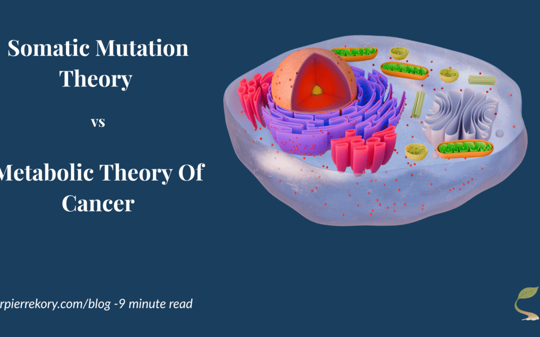
In this case, the wrong place is the nucleus. And the incorrect framework is the Somatic Mutation Theory of cancer, which is the consensus model that has guided oncology research and treatment for over 70 years. Evidence that overturned it has been sitting in the scientific literature for fifteen years, largely ignored by the institutions that fund cancer research and the oncologists who treat patients.

At Leading Edge Clinic, we have built a complementary cancer care program around the framework that replaced it: the Metabolic Theory of Cancer. This post explains what that theory is, why the evidence supports it, and what it means for how cancer should be treated, including the role of repurposed drugs and metabolic interventions that most oncologists are still not discussing with their patients.


Two Theories of Cancer: A Fundamental Disagreement

To understand why the Metabolic Theory of Cancer matters, you first need to understand what it is competing against.

The Somatic Mutation Theory (SMT) holds that cancer arises from the accumulation of genetic mutations in the DNA contained within the nucleus of a cell. Carcinogens damage genes, specifically tumor suppressor genes and tumor promoter genes, and these mutations drive the cell to acquire the eight hallmarks of cancer identified by Hanahan and Weinberg: uncontrolled growth, evasion of cell death, immune evasion, the ability to spread, and critically, a reprogramming of energy metabolism.

The SMT has been the foundational, total, and guiding theory of cancer since the early 1970s. It is the basis for the entire field of “targeted therapy” drugs designed to counteract the products of specific genetic mutations.

The Metabolic Theory of Cancer (MTOC) holds something fundamentally different. It posits that cancer begins not with nuclear DNA damage but with damage to the mitochondria, the energy-producing organelles in the cell’s cytoplasm. When mitochondria are damaged, they lose the ability to use oxygen for energy production and are forced instead to rely almost entirely on glucose fermentation. This metabolic switch is known as the Warburg Effect, and was first identified by Nobel laureate Otto Warburg in 1927. This switch is then what sends distress signals to the nucleus, disrupting DNA repair mechanisms and activating oncogenes. Under the MTOC, nuclear mutations are a consequence of mitochondrial dysfunction, not its cause.

With this distinction, comes a fundamental difference in what you treat, how you treat it, and with what.

Side-by-side comparison of two competing theories of cancer origin. Left panel (gray, Somatic Mutation Theory): posits that carcinogens damage DNA, mutations drive the eight cancer hallmarks, and the treatment approach is to target mutations with drugs. Outcome noted: no founding mutation has been found and 700 targeted drugs produced zero cures. Right panel (teal, Metabolic Theory of Cancer): posits that carcinogens damage mitochondria, damaged mitochondria send signals to the nucleus, and nuclear mutations are downstream effects. Outcome noted: nuclear transfer experiments confirm mitochondria are the origin. Source: Seyfried 2012, Warburg 1927, nuclear transfer experiments.


The Failure of the Somatic Mutation Theory

The SMT ran into serious trouble the moment large-scale genomic sequencing became possible.

The Cancer Genome Atlas Project

In 2005, the Cancer Genome Atlas Project (TCGA) was launched. It was a massive, well-funded effort to sequence the genomes of cancer cells across multiple cancer types. The goal was to identify the mutations driving cancer, providing the targets for the next generation of therapies. Vogelstein, the most prominent SMT researcher of his era and the scientist who found that p53 mutations occur in more than 50% of cancers, was centrally involved.

What the TCGA found was not confirmation of the SMT. It was its systematic dismantling.

Across tumor types, the project found random mutations throughout the genome — no orderly progression of successive mutations as the theory predicted. Few previously unknown oncogenes were identified. No single “founding mutation” — a mutation that would need to be present in every cancer cell if the SMT were correct — was found. Mutations differed dramatically between patients with the same cancer type. Even within a single patient’s tumor, mutations varied widely between cells. In breast cancer, five tumors had no mutations at all while still behaving aggressively and cancerously.

The last finding is particularly damning. If nuclear mutations cause cancer, then every cancer cell, being a daughter of the original mutated cell, must carry that founding mutation. If you find cancer cells without any founding mutation, the Somatic Mutation Theory is dead. No founding mutation has been found to date.

In response, Vogelstein repeatedly modified the SMT to accommodate the contradictory data, eventually proposing twelve distinct biological systems that cancer could disrupt, which Travis Christofferson in his book Tripping Over the Truth described as “an ad hoc modification necessary to make a failed theory fit the data.” He eventually abandoned the study of cancer causes entirely, shifting his focus to diagnostics.

The field of targeted therapies (seven hundred agents tested in solid tumor patients over the past decade) produced zero drugs that prolonged survival by one year compared to conventional treatment. Zero. This is what you get when you design drugs against targets that are consequences rather than causes.


The Metabolic Theory: What Warburg Found and Why It Was Ignored

Otto Warburg discovered in 1927 that cancer cells, unlike normal cells, rely almost exclusively on glucose fermentation for energy, even in the presence of oxygen. He spent the rest of his life arguing this was not a curiosity but the central defining feature of a cancer cell. He won the Nobel Prize in 1931 for this discovery.

The Warburg Effect is, notably, the biological basis of PET scanning, which are the imaging technology that every oncologist uses to diagnose, stage, and monitor cancer. PET scans inject radio-labeled glucose into patients and identify cancer by the areas of unusually high glucose uptake. Every oncologist who orders a PET scan is implicitly acknowledging the Warburg Effect, that cancer cells are defined by their glucose dependence, while simultaneously telling patients that diet has no bearing on their cancer.

The question Warburg could never answer in his lifetime was why. Why do cancer cells rely on glucose even when oxygen is available?

Pedersen, Seyfried, and the Nuclear Transfer Experiments

The answer came from decades of work by Pete Pedersen, beginning in 1979, and was assembled into a coherent theory by Dr. Thomas Seyfried at Boston College in the early 2000s.

Pedersen discovered that cancer cells had structurally abnormal mitochondria. There were fewer of them, they were smaller, and they had missing membranes and abnormal protein content. He found that cancer cells expressed a specific, overactive form of hexokinase that both drove glucose consumption to its maximum and simultaneously inhibited apoptosis (programmed cell death). He established that carcinogens which better damaged mitochondria were better at causing cancer than those which damaged chromosomes.

Seyfried assembled these findings into a complete theory, grounded in nuclear transfer experiments from the 1980s that had been performed and then effectively ignored.

In these experiments, researchers took the nucleus of a cancer cell and transferred it into a normal cell from which the nucleus had been removed. If cancer was caused by nuclear mutations — as the SMT held — these reconstituted cells should have behaved cancerously. Instead, in study after study, they did not. The healthy mitochondria in the host cell appeared to silence the cancerous nuclear DNA.

The reverse experiment was equally decisive. When mitochondria from a cancer cell were transferred into a normal cell with a healthy nucleus, 97% of the resulting mice developed cancer.

The conclusion was unambiguous: damaged mitochondria, not mutated nuclei, are the proximate cause of cancer. The mutations found in cancer nuclei are downstream consequences of mitochondrial dysfunction, not its upstream cause.

Seyfried published his synthesis in 2012 in his book Cancer as a Metabolic Disease. He received standing ovations at medical conferences. The National Cancer Institute declined to meaningfully shift its research funding priorities.


How the Two Theories Fit Together

It’s important to be precise here, because the MTOC does not deny that nuclear mutations exist in cancer — they clearly do. What it argues is that these mutations are not the primary driver. They are consequences of mitochondrial dysfunction.

Damaged mitochondria send signals back to the nucleus (what Seyfried called the “retrograde response”) that disrupt DNA repair mechanisms and activate oncogenes. The mutations observed in cancer cells arise because the cellular machinery for monitoring and correcting DNA errors has been compromised by the initial mitochondrial insult.

This means that the SMT is not entirely wrong. It is, as Seyfried has argued, incomplete. The mutations are real. They are just not the starting point. And designing cancer therapy exclusively around targeting those mutations,while ignoring the metabolic dysfunction that generated them, is precisely why targeted therapy has delivered such modest results.

The two theories can and should coexist in clinical practice. At Leading Edge Clinic, we do not advise patients to forego standard of care. We argue that standard of care should be complemented with metabolic interventions, because if metabolic dysfunction is primary, then metabolic intervention must be part of the treatment.


The Warburg Effect and the Ketogenic Diet

The most immediate clinical implication of the MTOC is dietary.

If cancer cells are defined by their dependence on glucose, and if normal cells can switch to burning ketone bodies derived from fat when glucose is unavailable  (while cancer cells generally cannot, because this requires functional mitochondria) then a ketogenic diet creates a metabolic environment that selectively disadvantages cancer cells while supporting normal ones.

Thomas Seyfried tested this in animal models and found that caloric restriction and ketogenic diets were anti-angiogenic, pro-apoptotic, anti-invasive, and restored normal aerobic metabolism. This pushes back on nearly every mechanism by which cancer cells sustain their cancerous behavior.

Published studies support beneficial impacts of ketogenic and caloric restriction approaches across multiple cancer types including breast, brain, colon, pancreas, lung, and prostate cancers. Studies also show that such dietary approaches reduce chemotherapy side effects and potentiate chemotherapy efficacy. Patients who combine ketogenic diet with radiation or chemotherapy appear to do better than with either alone.

When we ask our cancer patients what their oncologist told them about nutrition, the most common answer is: “whatever you like.” A PET scan that lights up because of glucose uptake, ordered by a clinician who tells their patient diet doesn’t matter, represents a profound disconnection between the diagnostic tool and the treatment philosophy.

Side-by-side cell diagram comparing normal cell metabolism to cancer cell metabolism. Left (normal cell): uses oxygen and functional mitochondria for oxidative phosphorylation, producing 36–38 ATP per glucose molecule, with CO₂ and water as byproducts. PET scan result: dim, indicating low glucose uptake. Right (cancer cell): mitochondria are damaged and unable to use oxygen; the cell ferments glucose even in the presence of oxygen (aerobic glycolysis), producing only 2 ATP and generating lactate. PET scan result: bright, indicating high glucose uptake. A callout at the bottom notes that PET scans identify cancer by the Warburg Effect, yet most oncologists do not recommend a ketogenic diet. Source: Warburg 1927, Seyfried 2012.


Repurposed Drugs: The Clinical Application of the MTOC

Metabolic dysfunction in cancer cells creates multiple therapeutic vulnerabilities that can be targeted by drugs already in clinical use for other indications. This is the foundation of the repurposed drug approach to cancer.

Dr. Paul Marik, co-founder of the FLCCC and Dr. Kory’s longtime colleague, conducted a comprehensive scoping review of nearly 1,500 scientific references on repurposed drugs and nutraceuticals in cancer treatment, compiled in his book Cancer Care. He graded the evidence for each compound, identifying seventeen with strong evidence, eight with weak evidence, and fifteen with insufficient evidence.

At Leading Edge Clinic, we work from the compounds with both strong published evidence bases, but also the backing of our own clinical experiences. The result is combinations of agents depending on cancer type and stage. The pathways we target with repurposed drug combinations include Hexokinase 2, p53, TGF-β, Wnt, Notch, PI3K/AKT, Hedgehog, IGF-1, and more. These are all metabolic or growth-signaling pathways that are disrupted in cancer cells.

What the Clinical Trials Show

The repurposed drug approach is not speculative. A growing body of clinical trial evidence, primarily from academic centers outside the United States, where practitioners are less financially constrained to standard of care, shows meaningful results.

CUSP9 (glioblastoma): Nine repurposed drugs added to standard of care. Phase 1 trial results: 30% of patients alive and disease-free at over four years post-treatment. Historical standard of care long-term survival: under 5–10%.

METRICS (glioblastoma): Four repurposed drugs — metformin, atorvastatin, mebendazole, and doxycycline — added to standard of care in 95 Stage IV patients. Two-year survival: 64% versus 26–28% in historical controls. Median survival: 27.1 months versus 14–15 months. 85% of patients tolerated all four drugs without difficulty.

CLOVA (glioblastoma): Four different repurposed drugs — cimetidine, lithium, olanzapine, and valproate — in seven patients with recurrent, chemotherapy-resistant GBM with the poorest predicted prognosis. Median overall survival after recurrence: 11.2 months versus 4.3–4.9 months in historical controls (p=0.004).

COMBAT (pediatric solid tumors): Celecoxib, vitamin D, fenofibrate, and retinoic acid alongside standard chemotherapy in 74 children with advanced, heavily pretreated tumors. In the high-grade sarcoma subgroup: median overall survival of 15.4 months versus 3.9 months in historical controls — nearly four times longer (p=0.001).

These are meaningful, consistent improvements in survival across multiple cancer types, in multiple independent research groups, with multiple different drug combinations. What they all share in common is the thread of targeting metabolic and growth pathways rather than specific genetic mutations.

The ReDO (Repurposing Drugs in Oncology) Project, a partnership between The AntiCancer Fund and Global Cures, has now identified 970 trials of repurposed drugs in cancer across 45 countries. There is real momentum and there are reall results behind this approach. The barrier to widespread scientific inquiry is primarily financial. Repurposed drugs are off-patent, so there is no commercial incentive to fund large trials, and pharmaceutical companies are not funding the research that would establish their efficacy.

Comparison table showing survival outcomes across four repurposed drug combination trials versus historical controls. CUSP9 (9 drugs, glioblastoma): 30% alive at 4+ years versus 5–10% in historical controls. METRICS (4 drugs including metformin, atorvastatin, mebendazole, doxycycline, glioblastoma Stage IV): 64% two-year survival and 27.1 months median survival versus 27% and 14.5 months in historical controls. CLOVA (4 drugs, recurrent GBM): 11.2 months median survival versus 4.6 months. COMBAT (4 drugs, pediatric solid tumors): 15.4 months median survival versus 3.9 months. A green summary bar notes that all four trials showed meaningful, consistent improvements across independent research groups, multiple drug combinations, and multiple cancer types. An amber bar notes that the barrier to wider adoption is that repurposed drugs are off-patent with no commercial funding incentive, and the ReDO project has identified 970 repurposed drug trials across 45 countries. Sources: CUSP9, METRICS, CLOVA, COMBAT trial publications.


Cancer Stem Cells: The Target Standard Therapy Misses

One of the most important, and most overlooked, aspects of why standard cancer therapies so often fail is the cancer stem cell problem.

Cancer stem cells (CSCs) are a small, resilient subset of tumor cells (comprising between 0.01% and 2% of a tumor) that drive growth, relapse, and metastasis. Unlike the rapidly dividing cancer cells that chemotherapy and radiation target, CSCs exhibit self-renewal, differentiation, and anti-apoptotic properties that make them resistant to conventional treatment.

More troublingly, there is published evidence that chemotherapy and radiation can actively promote CSC proliferation, meaning the treatments that shrink tumors can simultaneously stimulate the very cells most responsible for recurrence. There are currently no FDA-approved therapies specifically targeting cancer stem cells.

Repurposed drugs change this picture. Since the discovery of CSCs, extensive in vitro studies have identified affordable, accessible agents that inhibit CSC proliferation. At Leading Edge Clinic, we specifically target CSCs in our combination protocols, which may include agents such as metformin, ivermectin, mebendazole, doxycycline, curcumin, green tea extract, berberine, melatonin, statins, and vitamin D3. Of course, the combinations used are specific to individual patients, and this is not an exhausitve list.

Targeting cancer stem cells is a response to one of the most important reasons that standard therapies produce high initial response rates followed by relapse. If you shrink the tumor while stimulating the cells that will rebuild it, you have not solved the problem.


Our Approach at Leading Edge Clinic

Our position is explicit: we do not recommend that patients avoid standard of care oncology. What we offer is a complement to that care, a metabolic and repurposed drug framework layered alongside standard treatment, aimed at the mechanisms that standard treatment leaves unaddressed.

Our approach may include some combination of:

Metabolic intervention. Ketogenic and caloric restriction dietary protocols, individualized to the patient’s cancer type, treatment status, and physical condition. The Warburg Effect is real. A clinical approach that ignores dietary glucose is ignoring the biology.

Repurposed drug combinations. Drawn from the strongest evidence base, targeting multiple growth-driving pathways simultaneously. We do not use single agents. The evidence strongly favors combination approaches, which is why CUSP9, METRICS, and CLOVA used four to nine agents at once.

Cancer stem cell targeting. Specific agents selected for anti-CSC mechanisms, addressing the relapse problem that standard therapy systematically fails to address.

Observational study enrollment. Leading Edge Clinic is a study site for a prospective, IRB-approved observational study comparing outcomes in patients treated with complementary repurposed drug protocols versus standard of care alone. It is being conducted through the nonprofit Rebuild Medicine. This is the scientific infrastructure needed to generate the evidence that the field’s financial dynamics have prevented from being produced.


A Note on Realistic Expectations

What is this approach? What is it not?

The patients who respond most dramatically to repurposed drug and metabolic protocols are not a representative sample. Dramatic responses exist. Complete and near-complete resolution of metastatic disease exists, as documented in our own patient series. But, so do more modest responses, such as moderate shrinkage of tumors, or halting of disease progression. There are also, of course, patients who do not respond the way we are hoping they will. Welcome to cancer care. It is a moving target, and our approaches evolve with both our clinical experience, and emerging evidence.

We present our outcomes consecutively and transparently, not cherry-picked successes like some others in the space. Of five consecutive metastatic lung cancer patients treated at our clinic with combination repurposed drug protocols and ketogenic diets, four showed no cancer progression over the observation period. One achieved complete remission. Two of those patients (by their own choice, not our recommendation) received no conventional chemotherapy, radiation, or targeted therapies. These are meaningful results, and they should be interpreted in the context of what we know from the clinical trials above.

They are also preliminary. This is why the observational study matters. We need the data. We need to understand who responds and why, so that the clinical approach can be refined and the patients who are most likely to benefit can be identified.

What we can say with confidence is that the metabolic framework is scientifically sound, the repurposed drug evidence base is real and growing, and the gap between what standard oncology offers and what is available to patients willing to go further is substantial.

If you want to learn more about becoming a patient with our clinic, please use the following resources:


Leading Edge Clinic offers adjunctive cancer care alongside standard oncology, including repurposed drug protocols and metabolic interventions. We see cancer patients in all 50 states via telehealth. Leading Edge Clinic is a study site for the Rebuild Medicine observational study on repurposed drugs in cancer.

This article is for educational purposes and does not constitute medical advice. We strongly encourage all cancer patients to seek professional care for treatment.


Key References

Post-Exertional Malaise in Long COVID and Post-Vaccine Syndrome: Why “Exercise More” Is the Wrong Answer

Post-Exertional Malaise in Long COVID and Post-Vaccine Syndrome: Why “Exercise More” Is the Wrong Answer

Introduction: The Crash That Doesn’t Make Sense

A walk around the block, a few hours of focused work, a social event, or a workout you thought you were finally ready for. It didn’t feel excessive at the time.

Then, 12 to 72 hours later, something happened that has no parallel in “normal” human experience. Not tiredness. Not soreness. A complete systemic collapse, with fatigue so profound it feels neurological, brain fog that makes thinking feel like wading through quicksand, muscle pain, sensory sensitivity, and a return of every symptom you thought you’d been making progress on.

This is post-exertional malaise. And if you have Long COVID or Post-Vaccine Syndrome, you’ve almost certainly experienced it.

What you may not have received is an honest clinical explanation of what it actually is, why it happens, and why the treatment most clinicians prescribe — pushing through, gradually increasing activity, “deconditioning” management — is not just ineffective but genuinely harmful.

At Leading Edge Clinic, we treat PEM as one of the most mechanistically complex and most clinically important features of Long COVID and Post-Vaccine Syndrome. Getting it wrong doesn’t just stall recovery. It actively damages the biology you’re trying to repair.


What Post-Exertional Malaise Actually Is

PEM is formally defined as the worsening of symptoms following physical, mental, or cognitive exertion. It typically occurs with a delay of 12 to 48 hours after the triggering activity, and can last days to weeks.

The delay is important because it’s what makes PEM so consistently misunderstood by clinicians who aren’t familiar with it. If you crashed immediately after exertion, it would feel like an obvious cause-and-effect relationship. The delay means that by the time you feel the worst of it, you’ve already returned to baseline and the connection to what you did two days ago is no longer obvious. Patients are told their symptoms are unrelated to exercise. Sometimes they get an even worse explanation… “it’s all in your head”. Clinicians interpret the crash as a separate event rather than a predictable physiological consequence.

PEM is not deconditioning. It is not anxiety. It is definitely not a failure of effort or will – many of our patients were often “high achievers” prior to their illness. PEM is a specific, reproducible, biologically mediated response to exertion that occurs in a body with compromised cellular energy production, impaired microcirculation, and immune dysregulation. Even the mainstream research now makes this unambiguous.


The Biology: What Is Actually Happening During a PEM Crash

Mitochondrial Dysfunction at the Core

The most important mechanistic development in understanding PEM in Long COVID came from a series of muscle biopsy studies — most notably the 2024 landmark study published in Nature Communications by Appelman, Charlton, and colleagues at Amsterdam UMC, and the accompanying opinion article in Trends in Endocrinology and Metabolism by the same group.

What these studies found in the skeletal muscle tissue of Long COVID patients was as follows: intrinsic mitochondrial dysfunction, not just as a baseline impairment, but one that worsened measurably after exercise. Mitochondrial respiration (the process by which cells produce ATP) decreased significantly one day after maximal, PEM-inducing exercise. Markers of mitochondrial density dropped. The muscle showed acute tissue damage, focal necrosis, and intramuscular infiltration of immune cells that was not present before the exercise challenge.

Figure 5 from Appelman et al.,

This is not deconditioning. A deconditioned person’s muscles don’t necrotize after moderate exercise. What’s happening in Long COVID and Post-Vaccine Syndrome PEM is closer to a pathological injury response. The tissue cannot meet the energetic demand placed on it, and the attempt to do so causes measurable cellular damage.

The same research group also identified a shift in muscle fiber type, away from slow-twitch oxidative fibers and toward fast-twitch glycolytic fibers. This matters because oxidative fibers rely on efficient mitochondrial energy production for sustained activity. Glycolytic fibers burn energy quickly but produce lactate rather than sustained ATP. We have heard countless times from patients describing their PEM, some variation of, “it feels like my entire body is full of lactic acid. Like I’ve been poisoned.” A muscle that has shifted toward glycolytic metabolism will hit an energy wall faster, recover more slowly, and be more vulnerable to the kind of anaerobic overload that characterizes PEM.

The Microcirculation Problem

The mitochondrial dysfunction doesn’t occur in isolation. It is compounded by, and in part caused by, the endothelial dysfunction and microclotting that are central features of Long COVID.

When capillary beds are partially obstructed by amyloid fibrin microclots, oxygen delivery to working muscle tissue is impaired. The mitochondria of a Long COVID patient’s muscle cells may be operating in a relative hypoxic environment, even at rest. Under the increased oxygen demand of exercise, that deficit becomes significant. The tissue is asked to produce more energy precisely when its oxygen supply is most constrained.

A 2023 study in Acta Neuropathologica confirmed that Long COVID patients show capillary alterations and immune dysregulation in skeletal muscles. In other words, the physical infrastructure of oxygen delivery to muscle tissue is damaged. You cannot exercise your way out of damaged capillaries. Graded exercise therapy aimed at improving mitochondrial function in these patients is asking an engine with a clogged fuel line to run harder.

The Immune Activation Component

Exercise, even moderate exercise, triggers an inflammatory response in healthy individuals. The muscles release cytokines. Immune cells infiltrate tissue. This is part of normal adaptation and recovery. In healthy individuals, the process resolves and the tissue rebuilds stronger.

In Long COVID and Post-Vaccine Syndrome patients with already-elevated inflammatory signaling (already-elevated IL-6, already-activated mast cells, already-dysregulated immune responses), exercise delivers an additional inflammatory load onto a system with no reserve capacity to manage it. The immune activation triggered by exercise doesn’t resolve normally. It compounds the existing inflammatory state, triggering the systemic symptom cascade that characterizes a PEM crash.

Research published in PMC in 2025 confirmed that PEM in Long COVID is mediated by dysfunctions in both mitochondrial capacity and microcirculation, maintained by latent immune activation that conjointly impairs peripheral bioenergetics. This is not a psychological phenomenon. It is a coordinated biological failure at the cellular and vascular level.

The Cell Danger Response Connection

There is a deeper layer to PEM that connects to the Cell Danger Response framework we’ve written about previously.

When cells are in an active CDR state (as many Long COVID and Post-Vaccine Syndrome patients’ cells are), exertion represents exactly the kind of additional stressor that further activates the danger signaling cascade. ATP is released into the extracellular space as a danger signal. The purinergic signaling system, already dysregulated, interprets the metabolic stress of exercise as renewed threat. Rather than recovering normally, cells double down on the defensive metabolic state, suppressing normal energy production further, perpetuating the CDR, and deepening the very dysfunction that made exercise difficult in the first place.

This mechanism helps explain why PEM can leave patients worse off than before the triggering activity, not just temporarily, but potentially persistently if the CDR activation is severe enough to push cells further into the senescent cascade.

Side-by-side comparison of healthy muscle cell response to exercise (left, green) versus Long COVID muscle cell response to exercise (right, red). The healthy cell panel shows functional mitochondria with intact oxidative phosphorylation, open capillaries with adequate oxygen delivery, and normal recovery with tissue rebuilding stronger after exercise. The Long COVID cell panel shows impaired, fragmented mitochondria with ATP output collapsing under demand, a microclot blocking oxygen delivery to the cell, tissue hypoxia compounded by capillary obstruction, and post-exercise immune cell infiltration with focal necrosis and cell death. Both pathways lead to a bottom panel: the healthy pathway shows no crash, while the Long COVID pathway feeds into the PEM cascade — systemic crash, immune activation, CDR re-triggering, brain fog, and return of all symptoms — occurring 12–48 hours after activity. Sources: Appelman et al. Nature Communications 2024; Charlton et al. Trends in Endocrinology and Metabolism 2025; Aschman et al. Acta Neuropathologica Communications 2023.


The Graded Exercise Therapy Problem

Here is the clinical position that many uninformed clinicians are still operating from: Long COVID fatigue and exercise intolerance are primarily driven by deconditioning, and the solution is gradual, progressive increases in physical activity. This is also known as Graded exercise therapy, or GET.

This position is not just wrong, it is dangerous. It has been formally disputed by clinicians and researchers on the basis of the biological evidence, it was used in the ME/CFS context based on a deeply flawed trial (the PACE trial), and its application to Long COVID (and PACVS) patients with PEM has been specifically and publicly opposed by leading researchers including those at Amsterdam UMC, Mt. Sinai, and multiple European academic centers.

In January 2024, a letter published in Nature signed by Dutch, German, and Austrian investigators (joined by David Putrino of Mt. Sinai) stated explicitly: “We cannot agree with the recommendations for graded exercise therapy for people living with Long COVID who have post-exertional malaise.” The letter noted that the PACE trial, which is frequently cited in support of GET, has had its results called into question due to substantial protocol deviations and retrospective adjustment of recovery criteria. Improperly interpreted and manipulated study design and results? Where else have we seen that?

Professor Todd Davenport, a physical therapy researcher at the University of the Pacific, stated bluntly in 2024: “Any article published in 2024 citing the PACE trial as evidence of safety and efficacy of graded exercise should not be taken seriously.”

A review of exercise trials in Long COVID published in The Sick Times in late 2025 found that of 112 exercise-related trial registrations for Long COVID, fewer than 20% even mentioned PEM. Of those that did, two excluded participants with moderate-to-severe PEM and two excluded PEM participants entirely. The research community is studying exercise in Long COVID while systematically excluding the patients for whom exercise is most dangerous.

The deconditioning model fails for a simple reason: a deconditioned person’s skeletal muscle does not show focal necrosis, mitochondrial enzyme collapse, and immune cell infiltration after moderate exercise. That is what Long COVID patients’ muscle tissue shows. The biology is not consistent with deconditioning. It is consistent with a pathological injury response in tissue that lacks the cellular and vascular infrastructure to handle exertional demand.


What Appropriate PEM Management Actually Looks Like

If graded exercise therapy is the wrong answer, what is the right one? The honest clinical answer has two parts: understanding your energy envelope, and treating the underlying mechanisms that are causing PEM in the first place.

Pacing and the Energy Envelope

Pacing is not giving up. Many patients struggle to reconcile between their former selves, and their new physical constraints. But, pacing is a clinically rational strategy based on the biology of PEM. Specifically, the observation that there is a physiological threshold below which activity is tolerable and above which it triggers the pathological crash cascade.

The goal of pacing is to identify and stay within your energy envelope. Your energy envelope is the level of exertion your body can sustain without triggering post-exertional symptom worsening. This requires honest, often uncomfortable acceptance that your current energy envelope may be far smaller than your pre-illness capacity. Attempting to expand that envelope by pushing through symptoms (the instinct that works for normal fatigue, and has been socially built into many of our mindsets) actively damages the biology you’re trying to repair. It is easy to understand why patients may feel like they are going to “fall behind”, when adapting to this newfound situation.

Heart rate monitoring can be a useful pacing tool. Research suggests that staying below the anaerobic threshold, often approximated as 50–60% of age-predicted maximum heart rate in Long COVID patients, reduces the probability of triggering a crash. This is a lower threshold than most patients expect and lower than most clinicians prescribe.

Another extremely important and nuanced note… Cognitive and emotional exertion count. PEM is not exclusively triggered by physical activity. Mental effort, social interaction, sensory stimulation, and emotional stress can all trigger crashes in patients with significant PEM. This is consistent with: 1) the neurological and circulating contributors to PEM that the research has identified; and 2) the physiological facts that our brains account for 20% of our daily energy expenditure. All this means that pacing must encompass the full scope of daily exertion, not just physical exercise.

Line chart comparing two activity patterns over time against an exertion level axis. A dashed blue line shows pacing within the safe zone — staying below the Long COVID anaerobic threshold, which is marked lower on the chart than the healthy anaerobic threshold. A red solid line shows activity that crosses the Long COVID threshold, with a dotted return path showing the symptom trajectory peaking in a delayed PEM crash 12–48 hours after the activity stopped. The safe zone below the Long COVID threshold is shaded blue. The crash zone above it is shaded red. A callout box notes that a rising curve is inconsistent with residual antigen clearing. Sources: Charlton et al. Trends in Endocrinology and Metabolism 2025; Appelman et al. Nature Communications 2024.

Treating the Underlying Mechanisms

Pacing manages the symptom. It does not treat the cause. For Long COVID and Post-Vaccine Syndrome patients, the cause of PEM is the biological substrate described above: mitochondrial dysfunction, impaired microcirculation, immune dysregulation, and the ongoing CDR state and senescent cell burden.

Addressing microclotting. If capillary obstruction from amyloid fibrin microclots is impeding oxygen delivery to muscle tissue, reducing that obstruction through appropriate fibrinolytic support guided by clinical assessment (and where relevant, PAI-1 genotype) directly addresses one of the key contributors to PEM.

Mitochondrial support. The evidence for mitochondrial dysfunction in PEM is now well-established. Supporting mitochondrial function represents a rational clinical approach, though responses are individual and should be guided by a clinician familiar with this patient population.

Reducing the inflammatory environment. Since immune activation during exercise compounds the existing inflammatory state and drives PEM crashes, reducing the baseline inflammatory burden expands the effective energy envelope over time.

CDR-targeted approaches. For patients where the Cell Danger Response is a significant driver, interventions that support purinergic signaling normalization and allow cells to progress through the healing cycle are directly relevant to PEM. Reducing CDR activation reduces the hair-trigger sensitivity to exertion that characterizes severe PEM.

Careful sequencing. None of these interventions operates in isolation. PEM in Long COVID typically reflects multiple simultaneous mechanisms, and the clinical art is in identifying which mechanisms are dominant in a given patient and addressing them in the right sequence. A patient whose primary PEM driver is microclotting needs a different initial emphasis than a patient whose primary driver is MCAS-mediated immune activation after exercise.


What We Tell Our Patients

At Leading Edge Clinic, the practical guidance we give patients with significant PEM is this:

PEM is a real biological phenomenon with measurable cellular and vascular causes. It is not a symptom you can push through. Attempting to do so, without first addressing the underlying biology that is causing it, will not improve your condition… it will damage it.

However, there is good news. Your current energy envelope is not a permanent ceiling. It is a reflection of your current biological state. As we work on the underlying mechanisms (microclotting, immune dysregulation, CDR, mitochondrial function) your energy envelope will expand. We have seen it over and over. But that expansion has to come from biology improving, not from forcing activity in a system that isn’t ready for it.

This process takes time. It is non-linear. There will be weeks that feel like setbacks. Honest pacing and the discipline to stay within your current envelope even when you feel temporarily better is one of the most important things you can do to protect your recovery trajectory.

Recovery from PEM-dominated Long COVID is one of the slower trajectories we manage, and one of the most satisfying when it happens. Patients who have been bed-bound or housebound for years have regained meaningful function. But that recovery almost always required first stopping the cycle of crash and partial recovery that graded exercise therapy perpetuates, and replacing it with intelligent, mechanistically-informed clinical management.

If you’re interested in receiving care for your PEM, you can register here.


Leading Edge Clinic specializes in Long COVID, Post-Vaccine Syndrome, and complex post-infectious illness. Dr. Pierre Kory, Scott Marsland, FNP-C, and the rest of our clinical team have treated more than 3,500 patients, many who suffer with PEM. We see patients in all 50 states via telehealth.

This article is for educational purposes and does not constitute medical advice.


Key References

Why COVID Reinfection Matters More Than You Think — Especially Now

Why COVID Reinfection Matters More Than You Think — Especially Now

Introduction: The Wrong Question About Reinfection

Every time a new COVID variant emerges, the public health conversation follows the same script. Is it more severe? How many mutations does it have? Will the current vaccine protect against it (hah!)? Are hospitalizations rising?

For people with Long COVID, Post-Vaccine Syndrome, or any history of spike protein-related illness, they are almost entirely the wrong questions.

The correct question isn’t whether the COVID, or the latest variant Cicada variant (BA.3.2), causes more severe acute illness than previous strains. The correct question is: what does each additional COVID exposure do to someone who already has persistent spike protein, ongoing cellular senescence, and an immune system that never fully resolved the first round?

The answer, supported by emerging research from scientists at the Vedicinals group and consistent with what we observe clinically, is deeply concerning. It is also largely absent from mainstream COVID coverage.

This post explains why reinfection matters in a way that the standard messaging doesn’t capture, what the Cicada variant means specifically for this patient population, and what can be done to reduce the biological cost of repeated exposure.


What the Cicada Variant Actually Is

As of early April 2026, BA.3.2 has been detected in more than half of US states, according to the CDC. The WHO classified BA.3.2 as a variant under monitoring in December 2025, citing the variant’s many mutations and substantial antibody escape.

What makes Cicada notable is not its acute severity, but its spike protein. Compared to the current predominant strains of SARS-CoV-2, BA.3.2 carries 70 to 75 genetic changes in its spike protein. Of course, we alredy know the spike protein drives fibrinogen misfolding, mast cell activation, endothelial dysfunction, microclotting, and cellular senescence in Long COVID patients. But, Cicada’s mutations may help it evade antibodies. In a patient population where immune dysfunction is already significant, this is something Long COVID and Post-Vaccine Syndrome patients need to take note of for reasons explained later in this post.

Importantly, early data suggests BA.3.2 does not cause more severe acute disease than recent variants.

For the general public, that assessment is probably reassuring. For patients with existing spike protein burden and senescent cell accumulation, it misses the point entirely.


The Framework That Changes Everything: Spike Persistence and the Senescence Cascade

To understand why reinfection matters so much for this patient population, you need to understand a mechanistic framework that is only now beginning to enter the scientific literature, and that we at Leading Edge Clinic have been treating clinically.

A recently published paper from researchers at the Vedicinals group and collaborating institutions advances a hypothesis that brings together two phenomena that have been observed separately but rarely connected with sufficient clinical clarity – persistent spike protein production and progressive cellular senescence.

The key insight is this: Long COVID may not be primarily a problem of “how much spike is left over” from an infection. It may be a problem of ongoing spike production from a small population of cells that never stopped making it.

The Persistent Producer Cell

The paper proposes that a small number of cells, potentially harboring viral RNA in protected intracellular compartments called double-membrane vesicles, or having integrated spike-encoding genetic material, continue to produce and release spike protein long after the acute infection resolves. These “producer cells” generate modest amounts of spike individually, but their output is continuous, and it accumulates in tissues over time.

This would explain a striking observation from a high-volume European Long COVID diagnostic laboratory: spike protein detectability in Long COVID patients rose from roughly 30–40% in 2024, to approximately 75% in the first three quarters of 2025, to 96.5% by the fourth quarter of 2025. Under a simple “leftover antigen slowly clearing” model, you would expect a declining curve. A rising curve points toward ongoing production – an active source term, not residual debris from a resolved infection.

Line chart showing observed spike protein detectability in Long COVID patients across three timepoints: approximately 30–40% in 2024, approximately 75% in Q1–Q3 2025, and 96.5% in Q4 2025. The observed data is shown as a rising red line with labeled data points. A dashed gray line shows the expected trajectory under a clearance model, which would predict declining detection over time. An annotation box highlights that a rising curve is consistent with ongoing spike production rather than residual antigen slowly clearing. Source: Gerlach, Baig et al. 2025, Vedicinals Group, ELISA-based European Long COVID clinic data.

How Spike Spreads Beyond the Original Source

The paper outlines several mechanisms by which spike from a small number of producer cells can reach a vastly larger number of healthy cells:

Extracellular vesicles (exosomes). Spike protein packaged inside exosomes can evade antibody neutralization. Antibodies can’t bind to what they can’t reach. These vesicles carry spike through circulation and into tissues, delivering it to cells that never encountered the virus directly. These are the same vesicles by which proposed spike protein shedding events occur.

Syncytia formation. When spike-expressing cells contact cells with ACE2 receptors, they can fuse, creating multinucleated structures. Each fusion event effectively transfers spike-producing capacity to multiple adjacent cells simultaneously. What does this mean? It means a single producer cell can transfect 5–20 neighbors in one fusion event.

Tunneling nanotubes (TNTs). Cells can form thin, direct membrane bridges to neighboring cells through which spike protein, vesicles, and potentially viral RNA can transit — entirely shielded from antibody neutralization. A spike-producing cell may maintain TNT connections with 5–50 neighboring cells at once.

The result is progressive tissue saturation: a small upstream source driving a disproportionately large downstream burden.

The Senescence Cascade

The senescence cascade is where the model becomes clinically relevant.

As cells accumulate spike protein intracellularly – whether through exosomal uptake, syncytia fusion, or TNT transfer – they experience proteostatic stress, ER stress, and DNA-damage-response signaling. Once these signals cross a threshold, the cell enters a state of irreversible growth arrest: cellular senescence. It stops dividing, resists programmed cell death, and begins producing a continuous stream of pro-inflammatory signals collectively called the SASP — the senescence-associated secretory phenotype.

But the most important feature of senescent cells is that their SASP is contagious.

SASP factors — IL-6, IL-8, IL-1β, TNF-family molecules, matrix metalloproteinases — induce senescence in neighboring cells that may contain no measurable spike antigen themselves. This bystander or paracrine senescence means the lesion expands far beyond the originally spike-exposed population. Cells that never encountered spike become senescent because they were next to cells that did.

This creates a self-amplifying cascade that the paper describes precisely: the disease can transition from a spike-driven initiation phase to a senescence-dominant maintenance phase. In this case, the symptom burden becomes partially decoupled from measurable spike load. Patients remain severely symptomatic even when standard tests don’t detect viral material, because the biology has “handed off” from an antigenic driver to a self-sustaining cellular program.

This is consistent with what we observe clinically in Long COVID patients who have been ill for two, three, or four years. It is also consistent with why therapies aimed purely at viral clearance often underperform in established disease.

Vertical flowchart showing five sequential stages of the senescence cascade, each in a labeled box with arrows connecting downward. Stage 1 (coral): Persistent producer cell — DMV-protected viral RNA, ongoing spike synthesis. Stage 2 (coral): Spike released into circulation — free spike, exosomal cargo, syncytia, tunneling nanotubes. Stage 3 (purple): Primary senescence — spike uptake leading to ER stress and p16/p21 arrest. Stage 4 (purple): SASP activation — IL-6, IL-8, IL-1β, TNF, and MMPs secreted. Stage 5 (red): Self-sustaining inflammatory state — spike-negative cells perpetuate SASP, disease burden outlasts antigen detection. Callout boxes on the sides note antibody escape via exosomal shielding, irreversibility of senescence, paracrine spread to neighboring cells, and reinfection layering onto this base. A dashed loop arrow on the left shows the self-amplifying feedback cycle. Source: Gerlach, Baig et al., Vedicinals Group 2025.


Why Each Reinfection Layers Onto This Foundation

Each additional COVID exposure, whether from Cicada, any other variant, or a future strain, adds a new round of spike protein input to a system that is already struggling with ongoing production, progressive tissue saturation, and expanding senescent cell burden.

It replenishes the upstream source. A reinfection doesn’t just cause acute illness and then clear. It potentially seeds new producer cells, adds to the extracellular vesicle pool carrying spike, and re-exposes tissues that may have been recovering toward a threshold. The spike positivity data from the Vedicinals group’s European laboratory is consistent with this: a population of Long COVID patients showing rising, not declining, spike detection over time. Reinfections accelerate what was already an accumulating burden.

It compounds the senescence burden. Each new wave of spike-driven senescence induction is additive. Primary senescence from new spike exposure layers onto existing senescent cell populations. The paracrine cascade expands into new tissue territory. Patients who were at the margin of clinical stability (still functioning, managing their condition) can cross a tipping point following reinfection into significantly more impaired states.

The Cicada variant’s immune escape makes it more likely to establish a productive reservoir. For a Long COVID patient whose immune system is already dysregulated and whose neutralizing antibody response may be quantitatively or qualitatively impaired, a variant with enhanced antibody escape has a higher probability of establishing persistent producer cells rather than being rapidly cleared.

The spike protein variant may matter. The Vedicinals paper notes that earlier variants, including Omicron, appear to induce higher p16 and p21 expression (markers of cellular senescence) than ancestral strains, despite causing less severe acute illness. Cicada’s 70–75 spike mutations represent a distinct protein configuration. Whether this configuration affects senescence-induction kinetics is not yet established, but the direction of the existing data (that newer variants may be more senescence-inducing despite less acute severity) is something this patient population needs to take seriously.

The acute illness is not the danger. The downstream biology is.

This is the core message that is absent from every piece of mainstream Cicada variant coverage. The question “is it more severe?” is answered by hospitalization rates and ICU admissions. It does not capture what happens over the following 6, 12, or 18 months in someone already carrying significant senescent cell burden. It does not capture the cumulative cost of each reinfection to a biological system that has been running in a state of chronic dysregulation.

Stacked bar diagram showing cumulative senescence burden increasing with each reinfection event at three timepoints for the same patient. At the first timepoint (initial infection), a baseline senescence layer is established. At the second timepoint, a reinfection 1 layer and reinfection 2 layer are stacked on top. At the third timepoint, a Cicada exposure layer is added, pushing the total burden above a marked tipping point threshold shown as a dashed red horizontal line. To the right of the tipping point, a bordered box labeled


The Role of Senolytics: Reducing the Cost of Repeat Exposure

If the senescence cascade model is correct, and the evidence is increasingly consistent with it, then one of the most important things a Long COVID patient can do in the context of ongoing variant circulation is actively work to reduce their existing senescent cell burden. This reduces the foundation onto which a new exposure would layer, and it may limit the propagation of any new senescence cascade.

This is not a new clinical concept for us. Senolytic interventions (agents that selectively clear senescent cells) have been part of our treatment framework for Long COVID and Post-Vaccine Syndrome.

Some (but not all) clinically relevant therapies in this context include:

Intermittent fasting and autophagy promotion. The body’s cellular recycling program, autophagy, is one of the primary endogenous mechanisms for clearing dysfunctional cellular components. Caloric restriction and intermittent fasting protocols can meaningfully upregulate autophagy, and this can complement other senolytic approaches. However, as we’ve noted in our MCAS content, fasting protocols in Long COVID patients require clinical judgment. Not every patient can tolerate aggressive fasting, and the approach needs to be sequenced appropriately with other interventions.

Senomorphics — reducing SASP without clearing cells. For patients where aggressive senolytic dosing is not appropriate, senomorphic agents — those that reduce SASP output without necessarily clearing the senescent cells — can limit the paracrine propagation of the cascade. Low-dose rapamycin and metformin both have evidence in this area, and both are already used in relevant clinical contexts at our practice.

A 2021 study published in Science by Camell and colleagues provided direct evidence that senolytics reduce coronavirus-related mortality in aged mice, specifically by clearing the senescent cell burden that amplified the inflammatory response to viral infection. While this was in the context of acute infection rather than chronic Long COVID, the mechanism is directly relevant. Reducing pre-existing senescent cell burden before or after a new exposure limits how much the SASP-driven inflammatory cascade can amplify in response.

This is a clinically actionable implication. Patients with established Long COVID who maintain an ongoing senolytic protocol are not only treating their current disease, they are reducing the biological cost of the next inevitable exposure.


What This Means Practically for Long COVID and PACVS Patients Right Now

The COVID variant of the week is circulating in a population of Long COVID patients who are already carrying spike protein burden, established senescent cell populations, and a SASP-driven inflammatory environment.

The practical implications for this population are:

Reinfection prevention matters more for you than for the general population. Your risk includes compounding senescence, new producer cell seeding, potential tipping-point transitions in disease severity.

Senolytic maintenance is relevant timing. For patients already on senolytic protocols, ensuring adequate maintenance dosing during a period of active variant circulation is clinically sensible. For patients who have not yet incorporated senolytics into their treatment, this is a reasonable moment to discuss it with your provider.

Acute COVID treatment matters differently for you. If you do develop an acute Cicada infection, early intervention with antiviral therapy is relevant not primarily to prevent severe acute illness, but to limit the duration and magnitude of spike protein production, and therefore the new producer cell burden established during the infection. Shorter, lower-severity infection means less spike, means less new senescence induction.

The spike protein clearance and senolytic work you do now is investment against future exposures. Reducing your current spike burden and your current senescent cell load is not just about feeling better today, it is about building a lower biological baseline from which any future reinfection would cascade. This is why we treat Long COVID not as a single-point intervention but as an ongoing, evolving clinical relationship.


Conclusion: The Real Risk of Reinfection

The Cicada variant is being reported as “not more severe”. For the acute phase, that is accurate. But for the Long COVID and Post-Vaccine Syndrome patient, the frame of acute severity is inadequate.

The real risk of reinfection is not hospitalization. The real risk is what each additional exposure does to a biological system already operating under chronic spike pressure and expanding senescent cell burden. Each reinfection is not a reset. It is an addition to a running total that has consequences that unfold over months and years.

The senescence cascade hypothesis advanced by researchers at the Vedicinals group provides a mechanistic vocabulary for why patients experience progressive worsening over time despite no acute event. It explains why Long COVID symptoms can outlast any measurable marker of infection. And it clarifies what is at stake in the context of ongoing variant circulation.

This is the conversation that needs to be happening with Long COVID patients right now. At Leading Edge Clinic, it is the conversation we are having.

REGISTER AS A PATIENT → https://drpierrekorypatientportal.md-hq.com/registration


Leading Edge Clinic specializes in Long COVID, Post-Vaccine Syndrome, and complex post-infectious illness. Our providers treat patients across all 50 states via telehealth.

This article is for educational purposes and does not constitute medical advice.


Key References

  • Gerlach J, Baig AM, et al. Persistent Spike Protein Production and Progressive Tissue Saturation in Long COVID: Novel Hypothesis for a Senescence Cascade. Vedicinals Group / Health-Shield. 2025. [Preprint]
  • Camell CD, Yousefzadeh MJ, Zhu Y, et al. Senolytics reduce coronavirus-related mortality in old mice. Science. 2021;373:eabe4832. https://doi.org/10.1126/science.abe4832
  • Patterson BK, et al. Persistence of SARS-CoV-2 S1 protein in CD16+ monocytes in PASC up to 15 months post-infection. Front Immunol. 2021;12:746021. https://doi.org/10.3389/fimmu.2021.746021
  • Meyer K, et al. SARS-CoV-2 spike protein induces paracrine senescence and leukocyte adhesion in endothelial cells. J Virol. 2021;95:e00794-21. https://doi.org/10.1128/JVI.00794-21
  • Tsuji S, et al. SARS-CoV-2 infection triggers paracrine senescence and a sustained senescence-associated inflammatory response. Nat Aging. 2022. https://doi.org/10.1038/s43587-022-00170-7
  • Gorgoulis V, et al. Cellular senescence: defining a path forward. Cell. 2019;179(4):813–827. https://doi.org/10.1016/j.cell.2019.10.005
  • Acosta JC, et al. A complex secretory program orchestrated by the inflammasome controls paracrine senescence. Nat Cell Biol. 2013;15(12):1524–1535. https://doi.org/10.1038/ncb2871
  • CDC. BA.3.2 (Cicada) Variant Monitoring Report. April 2026. https://www.cdc.gov/covid/variants/
Microclotting in Long COVID and Post-Vaccine Syndrome: How Your Genetics Impact Treatment

Microclotting in Long COVID and Post-Vaccine Syndrome: How Your Genetics Impact Treatment

Introduction: The Clotting Problem Most Doctors Aren’t Testing For

Many Long COVID and Post-Vaccine Syndrome patients know their symptoms. Fatigue that doesn’t resolve with rest. Brain fog that feels like you’re wading through muck just to formulate a coherent thought. Breathlessness that appears without warning. Chest tightness that comes and goes. Post-exertional crashes that wipe out any attempt at normal activity.

What most patients, and most clinicians, have come to know is that for a significant proportion of these patients, the blood itself is part of the problem.

Not in the dramatic, visible way that shows up on imaging. In a quieter, more insidious way: tiny, abnormal clots forming throughout the microvasculature, reducing oxygen delivery to tissues, trapping inflammatory molecules, and physically obstructing circulation in capillaries too small to show up on any standard scan. Amyloid fibrin microclots are not a novel issue. But, with spike protein, they are more significant than ever before. Not a single patient of ours comes to us with low levels of microclotting.

A critical aspect of microclotting we will explore in this article: a genetic variant that approximately 25–30% of the general population carries means some patients are dramatically more prone to forming these clots, and dramatically less able to clear them, than others.

Understanding this mechanism, and how your genetics interact with it, changes what treatment should look like. At Leading Edge Clinic, it’s something we assess in patients where microclotting is suspected as a significant driver. This post explains the underlying science and the clinical implications.


What Are Fibrin Amyloid Microclots?

Normal blood clotting is a tightly regulated process. When a vessel is injured, a soluble protein circulating in plasma called fibrinogen is converted to fibrin, forming a mesh-like clot that stops bleeding. Once the injury heals, an enzyme called plasmin dissolves the fibrin through a process called fibrinolysis. The clot clears. Normal circulation resumes.

In Long COVID and Post-Vaccine Syndrome, this process goes wrong in a specific and well-documented way.

Research pioneered by Professor Etheresia Pretorius at Stellenbosch University and Professor Douglas Kell at the University of Liverpool, beginning in 2021 and now confirmed across multiple independent research groups, has established that the spike protein of SARS-CoV-2 can trigger fibrinogen to misfold into an abnormal, amyloid-like form. These fibrinaloid microclots, the term used in the published literature, have structural properties that make them fundamentally different from normal clots.

Most critically: they resist normal fibrinolysis. The body’s standard clot-clearing machinery, plasmin, cannot effectively break them down.

Side-by-side diagram comparing normal fibrin clot dissolution with amyloid fibrin microclot persistence. Left panel (normal clot): fibrinogen is converted to normal fibrin mesh by thrombin, plasmin penetrates and cleaves the fibrin, and the clot is cleared with normal circulation restored. Right panel (amyloid microclot): spike protein triggers fibrinogen misfolding into dense amyloid fibrin, plasmin is blocked from penetrating the structure, PAI-1 overexpression in 4G/4G genotype further suppresses fibrinolysis, and the microclot persists — causing capillary obstruction, tissue hypoxia, and trapped inflammation.

What Makes These Microclots Different

Normal fibrin clots form a loose mesh that plasmin can penetrate and degrade. Amyloid fibrin microclots are densely compacted, beta-sheet rich structures. This is the same structural architecture seen in amyloid proteins associated with Alzheimer’s and Parkinson’s diseases. Plasmin can penetrate normal clots. It cannot efficiently dissolve amyloid fibrin.

Beyond their structural resistance, these microclots also trap inflammatory molecules within their matrix. Proteomics analysis by the Pretorius and Kell groups found that Long COVID microclots contain elevated levels of pro-inflammatory proteins, complement activation markers, and von Willebrand factor — creating what amounts to mobile packages of inflammatory material that continuously activate the immune system wherever they circulate.

A 2022 landmark paper by Kell, Laubscher, and Pretorius in the Biochemical Journal formally established microclots as a central driver of Long COVID pathology, noting that these structures persist in the plasma of Long COVID patients even when they are not in an active clotting event. They circulate freely, obstructing capillaries, and sustaining a chronic inflammatory state.

More recent research published in the Journal of Medical Virology (2025) confirmed that microclots in Long COVID patients are structurally associated with neutrophil extracellular traps (NETs), providing another mechanism by which they perpetuate thromboinflammation and immune dysregulation.

The Symptoms Microclotting Produces

The capillary bed is where oxygen, nutrients, and cellular waste products are exchanged between blood and tissues. When microclots obstruct these vessels, the consequences are predictable:

  • Fatigue and post-exertional malaise: Reduced oxygen delivery to muscles and mitochondria
  • Brain fog and cognitive dysfunction: Impaired cerebral microcirculation
  • Breathlessness and exercise intolerance: Reduced pulmonary capillary perfusion
  • Small fiber neuropathy and tingling: Nerve tissue hypoxia from microvascular obstruction
  • Chest discomfort: Myocardial microvascular involvement
  • Temperature dysregulation: Peripheral microcirculatory dysfunction

These are among the most commonly reported and most treatment-resistant symptoms in Long COVID. For patients where microclotting is a significant driver, failing to address it means failing to address a foundational cause of their persistent symptoms. Of course, these symptoms can have other potential driving factors that also must be addressed, such as MCAS, chronic Cell Danger Response, POTS, and more.


PAI-1: The Body’s Clot-Clearing Brake

To understand why some patients are far more vulnerable to persistent microclotting than others, you need to understand a protein called Plasminogen Activator Inhibitor-1, or PAI-1.

PAI-1 is the primary regulator of fibrinolysis, the clot-clearing process. Its job is to inhibit the enzymes (tissue plasminogen activator, or tPA, and urokinase plasminogen activator, or uPA) that convert plasminogen into plasmin, the enzyme that dissolves fibrin. In other words, PAI-1 is the brake on clot dissolution.

This braking function is necessary. The body doesn’t want clots dissolving prematurely when they’re serving a purpose (ie: stopping bleeding). But in a context where amyloid microclots are forming continuously and need to be cleared as quickly as possible, excessive PAI-1 activity is a serious problem. It keeps the brake partially applied when you need full fibrinolytic capacity.

And here’s where genetics becomes directly clinically relevant.


The PAI-1 4G/5G Polymorphism: A Genetic Modifier of Microclotting Risk

In the promoter region of the SERPINE1 gene, the gene that encodes PAI-1, there is a well-characterized genetic variant called the 4G/5G polymorphism. This refers to a single position in the DNA sequence where individuals carry either four consecutive guanosine bases (4G) or five (5G).

This small difference has significant functional consequences for how much PAI-1 your cells produce.

The Three Genotypes

The 4G/5G polymorphism produces three possible genotypes:

4G/4G (homozygous 4G): Both copies of the gene carry the 4G allele. The 5G allele has an additional transcriptional repressor binding site that reduces PAI-1 gene expression — the 4G allele lacks this site. Carrying two 4G alleles means higher baseline PAI-1 production, suppressed fibrinolysis, and significantly elevated thrombosis risk. Under inflammatory conditions, including COVID-19 infection and spike protein exposure, PAI-1 production in 4G/4G individuals ramps up further and is more difficult to suppress.

4G/5G (heterozygous): One copy of each allele. PAI-1 levels are intermediate. Research shows that inflammatory signals like IL-1β still enhance PAI-1 production in 4G/5G endothelial cells, though less dramatically than in 4G/4G individuals.

5G/5G (homozygous 5G): Both copies carry the 5G allele. Lowest baseline PAI-1 production, most active fibrinolysis. However, and this is a crucial nuance, 5G/5G individuals have a different risk profile in COVID-19 contexts. With the fibrinolytic brake released, these patients can develop overactive fibrinolysis and inflammation-driven endothelial dysfunction through a different mechanism.

A 2024 study published in Frontiers in Immunology by Yatsenko, Heissig, and colleagues at Juntendo University confirmed these distinct mechanistic profiles in COVID-19 patients, finding that 4G/4G individuals showed high circulating PAI-1 complexed with plasminogen activators, low plasmin levels, and NF-κB upregulation – a pattern of fibrinolytic shutdown under inflammatory conditions. The 5G/5G group showed the opposite: lower PAI-1, elevated free plasminogen activators, and a profile of inflammation-driven endothelial dysfunction.

Population Prevalence

The 4G allele is common. Population genetics research suggests approximately:

  • 25–30% of people carry the 4G/4G genotype
  • 50% carry the 4G/5G heterozygous genotype
  • 20–25% carry the 5G/5G genotype

This means roughly half to three-quarters of the general population carries at least one 4G allele — and among patients with severe, persistent Long COVID, this proportion may be even higher given the known interaction between the 4G allele and spike protein-driven inflammatory signaling.

Diagram showing the PAI-1 4G/5G polymorphism spectrum across three genotypes from left to right, ordered by increasing PAI-1 expression. 5G/5G genotype (approximately 20–25% of the population): lowest PAI-1 production, most active fibrinolysis, lower microclot burden, but risk of inflammation-driven endothelial dysfunction; treatment priority is anti-inflammatory first with fibrinolytics used cautiously. 4G/5G genotype (approximately 50% of the population): intermediate PAI-1 level that rises under inflammation, moderate microclot burden; treatment priority is moderate fibrinolytics such as nattokinase and sulodexide alongside anti-inflammatory support. 4G/4G genotype (approximately 25–30% of the population): highest PAI-1 production, NF-κB upregulation, severely suppressed plasmin activity, highest microclot burden; treatment priority is fibrinolytic-first using nattokinase for PAI-1 inhibition, lumbrokinase, and sulodexide. Source: Yatsenko et al., Frontiers in Immunology 2024.


Why This Matters Clinically: Two Different Problems, Two Different Approaches

The clinical significance of the 4G/5G polymorphism is not merely academic. It has direct implications for how microclotting should be treated in individual Long COVID and Post-Vaccine Syndrome patients, and why a uniform anticoagulation approach for all patients is inadequate.

The 4G/4G Patient: Fibrinolytic Suppression

For patients with the 4G/4G genotype, the central problem is that spike protein-driven inflammation severely suppresses fibrinolysis. PAI-1 rises under inflammatory conditions, plasmin activity is reduced, and the body’s capacity to clear microclots is significantly impaired.

In these patients, the therapeutic priority is: reduce PAI-1 activity and/or directly enhance fibrinolysis. This is where fibrinolytic enzymes – nattokinase, lumbrokinase – become particularly relevant. Both enzymes work through mechanisms that directly counter the 4G/4G problem.

Nattokinase is a serine protease derived from fermented soybeans (natto) that works through two complementary mechanisms: it directly cleaves fibrin, and it inactivates PAI-1 — the precise molecular target that is overexpressed in 4G/4G patients. Research published in the Journal of Agricultural and Food Chemistry demonstrated that nattokinase directly hydrolyzes PAI-1, increasing fibrinolytic activity. The combination of direct fibrin cleavage and PAI-1 inhibition makes it particularly well-suited to the 4G/4G mechanism.

Lumbrokinase, derived from earthworm species, operates through direct fibrinolytic action and plasminogen activator stimulation. Research by the PolyBio Research Foundation has initiated a clinical trial specifically examining lumbrokinase in Long COVID and ME/CFS, reflecting the growing clinical and mechanistic case for fibrinolytic enzymes in microclot-driven post-viral illness.

Beyond enzymes, sulodexide – a glycosaminoglycan with both anticoagulant and endothelial-protective properties – is used in our practice for patients with evidence of microclotting and endothelial involvement. Unlike systemic anticoagulants, sulodexide has a favorable safety profile for longer-term use and directly supports endothelial repair, which is important given that endothelial dysfunction is both a consequence of microclotting and a driver of further PAI-1 elevation.

The 5G/5G Patient: Inflammation-Driven Endothelial Dysfunction

For patients with the 5G/5G genotype, the problem is different. Fibrinolysis is not suppressed, but inflammation-driven endothelial dysfunction creates a prothrombotic state through other pathways, including elevated uPA and activated complement. These patients may be more prone to systemic inflammation and immune dysregulation than to pure fibrinolytic failure.

The 2024 Juntendo University study specifically identified 5G/5G patients as being at risk for inflammation-induced endothelial dysfunction with fibrinolytic overactivation, a phenotype where aggressive fibrinolytic therapy carries different risk considerations and where anti-inflammatory and endothelial-supportive strategies may be the more appropriate primary focus.

This is clinically significant: the 5G/5G patient who receives high-dose fibrinolytic enzymes without consideration of their genotype is receiving a treatment rationale designed for a different problem. Anticoagulation approach needs to be matched to mechanism, not applied uniformly.

The 4G/5G Patient: Intermediate Risk, Moderate Response

Heterozygous patients show an intermediate profile. The Juntendo study found that IL-1β still enhances PAI-1 production in 4G/5G endothelial cells, though less severely than in 4G/4G. These patients benefit from fibrinolytic support but may need less aggressive dosing and may respond well to lower-intensity anticoagulation combined with strong anti-inflammatory support.


Testing: What We Look For and When

Knowing a patient’s PAI-1 genotype is straightforward. It can be obtained through standard genetic testing including Labcorp’s PAI-1 4G/5G Polymorphism panel (test code 500309).

Beyond genotype, we also look at functional markers of clotting and fibrinolytic activity when clinically indicated:

  • PAI-1 functional activity levels (plasma)
  • D-dimer (marker of ongoing fibrin degradation — elevated in active microclotting)
  • Fibrinogen levels
  • Von Willebrand factor antigen (marker of endothelial activation)
  • Microclotting levels through fluorescence microscopy

Importantly, standard blood tests and imaging do not detect microclots. The specialized fluorescence microscopy techniques used in Pretorius’s laboratory research are not commercially available at just any lab. We are able to offer this testing to our patients.

This is an area where pattern recognition from clinical experience matters significantly. The presentation of a patient with 4G/4G genotype, elevated D-dimer, elevated fibrinogen, and symptoms strongly suggestive of microvascular obstruction tells a coherent story that guides a different treatment approach than a patient with 5G/5G genotype and primarily inflammatory, autonomic symptoms.


The Broader Picture: Microclotting Doesn’t Operate in Isolation

It’s important to place microclotting in the context of Long COVID’s full complexity. Persistent microclots are a significant mechanism for many patients, but they rarely operate alone. They interact with and compound other pathophysiological drivers:

Microclotting and Cell Danger Response: Tissue hypoxia from microvascular obstruction can itself activate and sustain the Cell Danger Response. Cells detect oxygen insufficiency as a threat and shift into the protective metabolic state we discussed in our CDR post. Addressing microclotting may be a necessary prerequisite to allowing the CDR to resolve in some patients.

Microclotting and POTS/Dysautonomia: Endothelial dysfunction from microclotting directly affects autonomic regulation of vascular tone. Many patients with post-COVID POTS have a vascular endothelial component to their dysautonomia that won’t fully resolve without addressing the underlying endothelial damage.

Microclotting and Neurological Symptoms: The 2025 Journal of Medical Virology study confirmed that microclots in Long COVID are structurally associated with NETs markers, including myeloperoxidase and neutrophil elastase, which can themselves cross the blood-brain barrier and contribute to neuroinflammation. Brain fog in these patients has a partially vascular etiology, not just neurological.

Microclotting and Senescent Cells: Chronic endothelial damage from persistent microclotting can itself drive cellular senescence in vascular endothelial cells, creating a feedback loop where senescent endothelial cells produce SASP-driven pro-inflammatory and pro-thrombotic signals that generate more clotting. This interaction is one reason why microclotting in some patients is difficult to resolve without simultaneously addressing cellular senescence.

The clinical implication is that microclotting treatment is rarely sufficient as a standalone intervention. It typically needs to occur in parallel with reducing the spike protein burden that is driving fibrinogen misfolding, addressing the inflammatory environment that elevates PAI-1, and supporting the endothelial repair that allows normal fibrinolytic function to resume.

Diagram showing how microclotting connects to four downstream mechanisms in Long COVID and Post-Vaccine Syndrome, all converging on persistent symptoms. Microclotting (center top) caused by amyloid fibrin and capillary obstruction drives two pathways: left, to cell danger response, where tissue hypoxia activates and sustains the CDR; right, to dysautonomia, where endothelial damage disrupts autonomic vascular tone. Cell danger response connects downward to cellular senescence, where endothelial damage drives senescent cell accumulation and a pro-thrombotic SASP feedback loop. Dysautonomia connects downward to neurological symptoms, driven by NETs crossing the blood-brain barrier and cerebral hypoperfusion. All four downstream nodes converge on a persistent symptoms box at the bottom, including fatigue, brain fog, post-exertional malaise, breathlessness, and neuropathy. A dashed feedback arrow from cellular senescence back toward the center indicates the pro-thrombotic loop that perpetuates further microclot formation. Root drivers — spike protein, PAI-1 4G/4G genotype, and endothelial injury — are labeled between the central node and the downstream pathways.


A Note on Safety and Clinical Oversight

Fibrinolytic therapy, whether enzymatic or pharmaceutical, requires appropriate clinical supervision. The primary risk is bleeding, and the probability of this risk increases meaningfully when fibrinolytic agents are combined with pharmaceutical anticoagulants (aspirin, clopidogrel, apixaban, warfarin, heparin) without careful monitoring.

This is not an argument against fibrinolytic treatment. We have treated over a thousand patients with triple anticoagulation therapy combined with enzymatic therapies. It is an argument for doing it with proper clinical oversight, appropriate dosing based on individual presentation and genotype, and awareness of the full medication picture. The patients who have the worst outcomes with DIY fibrinolytic protocols are typically those combining multiple agents without understanding their additive effects. Not only that, they are not dealing with the complete picture of spike protein injury.

At Leading Edge Clinic, anticoagulation approach is individualized to the patient’s genetic profile, symptom presentation, functional markers, and complete medication list. For some patients, nattokinase alone at appropriate dosing is the right starting point. For others, sulodexide plays a primary role. For a smaller number of patients with more significant microclotting burden and appropriate clinical indicators, pharmaceutical anticoagulation is warranted. There is no universal protocol.


Conclusion

Fibrin amyloid microclots represent one of the most mechanistically coherent and clinically important, yet most frequently missed, drivers of persistent Long COVID and Post-Vaccine Syndrome symptoms. They explain a cluster of symptoms (fatigue, brain fog, breathlessness, post-exertional malaise) that don’t respond to anti-inflammatories alone because the problem isn’t only inflammation. The problem is physical obstruction of microcirculation and ongoing thromboinflammation.

And the PAI-1 4G/5G polymorphism explains something that purely inflammation-focused frameworks can’t: why patients with similar spike protein exposure and similar inflammatory burdens have dramatically different microclotting trajectories. Your genetics determine how effectively your body can clear these abnormal clots, and they should determine how you treat them.

This is the kind of individualized, mechanism-informed clinical reasoning that drives our approach at Leading Edge Clinic. If you’re experiencing symptoms consistent with microclotting and you haven’t been evaluated for fibrinolytic capacity or PAI-1 genotype, that may be a meaningful gap in your care picture.

You can register here to become a patient.


Leading Edge Clinic specializes in Long COVID, Post-Vaccine Syndrome, and complex post-infectious illness. Our providers treat patients across all 50 states via telehealth. Initial evaluations are 60 minutes.

This article is for educational purposes and does not constitute medical advice. Fibrinolytic therapy carries clinical risks and should be undertaken only with appropriate medical supervision.


Key References

Why You’re Still Sick: What Most Long COVID Clinics Get Wrong

Why You’re Still Sick: What Most Long COVID Clinics Get Wrong

You’ve done everything right.

You went to your primary care doctor. They ran the standard panels, found nothing, and told you to rest. You pushed. Through endless research and self-advocacy, you found a Long COVID clinic – maybe a university program, maybe a telehealth practice, maybe an integrative specialist someone in your online support group recommended. You spent a significant amount of money on testing and extensive protocols,  waiting to get better.

You’re still sick.

If that describes your experience, this post is for you. Our goal is to honestly explain something that most Long COVID and Post-Vaccine Syndrome clinicians — even well-meaning, intelligent ones — fundamentally misunderstand about this condition.

At Leading Edge Clinic, we’ve treated more than 3,500 patients with Long COVID and Post-Vaccine Syndrome. Many of them came to us after failing at other practices, sometimes after spending $30,000 to $100,000 elsewhere. What we’ve observed over years of clinical work has shaped a different understanding of why standard approaches so often fall short.


The Core Problem: Long COVID Is Not One Disease

The most common clinical mistake we see, across conventional medicine, integrative medicine, and Long COVID specialty practices alike, is treating this condition as if it has a singular mechanism that responds to a singular approach (ie: dealing only with spike persistence, dealing only with immune dysfunction, looking only at gut health, etc…)

It doesn’t.

Long COVID and Post-Vaccine Syndrome are multi-system, multi-mechanism conditions that present differently in every patient and evolve over time. What’s driving your fatigue may not be the same as what’s driving the next patient’s. And critically, what’s driving your symptoms today may not be what’s driving them six months from now.

Like other historicaly post-viral and post-vaccine syndromes, this not a simple infection with a clean recovery arc. It is a dysregulated biological state, and it is very effcient at driving that dysregulated state — often involving the immune system, the nervous system, the vascular system, and the metabolic system simultaneously, in proportions that vary person to person and shift in response to treatment.

A clinician who approaches it with a fixed protocol, even a sophisticated one, is going to miss the mark for a significant portion of patients.


What Standard Approaches Miss

This is not an exhaustive list. It hits on some major systemic issues we don’t see others talking about, but these aren’t the only things we see other clinicians missing when a new patient comes to us that has tried elsewhere first.

1. They Don’t Account for Cell Danger Response

Basic diagram illustrating an optimal versus abnormal Cell Danger Response. The abnormal CDR shows ATP leakage of the cellular membraien

One of the most underappreciated mechanisms in Long COVID is something called the Cell Danger Response (CDR) — a concept developed by metabolic researcher Dr. Robert Naviaux.

The CDR is the body’s ancient, conserved response to threat. When cells detect danger, such as infection, toxin, or injury, they shift into a defensive metabolic state. Energy production is reduced, cellular communication slows down , and cells essentially go quiet to protect themselves.

This is adaptive in the short term. After an acute COVID infection, you want this response. It’s doing its job.

The problem is when the CDR doesn’t turn off. When the body’s threat detection system remains activated long after the initial danger has passed, you get persistent metabolic suppression. This manifests as fatigue that doesn’t resolve with rest, cognitive dysfunction that doesn’t respond to sleep, and a nervous system that stays in a low-grade defensive posture.

Most Long COVID clinicians are not testing for or thinking about CDR biology. Many deal with chronic inflammation and persistent spike protein, but never take that next step to address chronic CDR.

2. They Don’t Understand Senescent Cells

Diagram showing possible impacts of senescent cell accumulation, including: tissue reprogramming, osteoporosis, glaucoma, type 2 diabetes, neurodegeneration, immune dysregulation, cancer, tissue fibrosis, and impraied tissue repair

Cellular senescence, what some researchers call the “zombie cell” problem, is another mechanism that rarely appears in standard Long COVID treatment frameworks, although it may accidentally be addressed (albeit not thoroughly enough) through treatments with other prescribed purposes.

Senescent cells are cells that have stopped dividing but refuse to die. In the aftermath of a severe immune activation like COVID infection, the body can accumulate these cells in significant numbers. In fact, the spike protein seems to have be very skilled at producing senescent cells. These cells don’t do their normal jobs, but they also release a continuous stream of pro-inflammatory signaling molecules, the senescence-associated secretory phenotype (or SASP).

The result of senescent cell accumulation is a low-grade, chronic inflammatory state that can persist for years. Standard anti-inflammatory approaches don’t clear senescent cells. Senolytics, which are treatments that induce apoptosis in senescent cells, are rarely considered in clinical practice, let alone in Long COVID or Post-Vaccine Syndrome.

For patients where senescent cell burden is a significant contributor, which is all spike protein patients, treating everything else while ignoring this mechanism is like bailing water from a boat without fixing the leak.

3. They Apply Uniform Protocols to Non-Uniform Patients

We understand why this happens. Protocols are efficient. They can be standardized, taught, and scaled. If you’re running a practice that sees 50 Long COVID patients a week, a decision tree makes sense. Our practice model focuses on lower patient volumes for more personalized care and treatment. Why?

Long COVID patients are not uniform. The neurotransmitter profile of one patient – the specific pattern of catecholamine deficiency, glutamate dysregulation, and kynurenine pathway disruption – may look nothing like the next patient’s. The autoimmune burden, the degree of microclotting, the level of residual spike protein activity, the autonomic dysfunction pattern, may all vary enormously from patient to patient.

Our practice partner Scott Marsland, FNP-C, published a detailed case study that illustrates this complexity in just one patient, examining neurotransmitter changes across time and in response to specific interventions. The data showed 13 of 27 measured neurotransmitters were outside optimal range at baseline. The specific pattern of elevated glutamate suggesting excitotoxicity, paradoxically elevated serotonin alongside low tryptophan, and depleted catecholamines required a tailored response, not a standard protocol. And the picture changed meaningfully over seven months, requiring ongoing adjustment.

That is what individualized Long COVID care actually looks like.

4. They Over-Test and Under-Treat

There are particular kind of specialty practices that run $2,000–$5,000 in laboratory panels before doing much of anything. The testing is framed as “comprehensive” and “data-driven.” It feels thorough. As humans, we want to latch onto something concrete, like lab results. It gives us something to work at, and improve upon.

But here’s the honest clinical reality: in most Long COVID and PACVS patients, extensive testing rarely changes the initial treatment approach. The patterns we see across 3,500+ cases are recognizable. The likely mechanisms become apparent through careful history, symptom characterization, and clinical pattern recognition long before any lab results return.

Excessive testing delays treatment. It costs patients money they could have spent on interventions. And it creates an illusion of certainty in a condition that requires the intellectual honesty to acknowledge: we are treating this by thoughtful trial, observation, and adjustment. Running a panel and following a chart does not work for complex conditions like Long Covid and Post-Vaccine Syndrome. One day might turn up a normal lab result, and then many abnormalities the next testing period. Patients end up chasing ghosts.

And, by the way, if and when a $4,000 testing panel arrives that meaningfully guides pin-pointed treatment decisions tha deliver results, we will be the first ones to come back and edit this post.

5. They Treat the First Layer and Stop

Long COVID recovery, when it happens, typically unfolds across 9 to 18 months. Sometimes longer. The condition does not resolve in a single treatment arc, but requires ongoing attention to how the patient’s picture is shifting.

A treatment that was appropriate in month two may need modification by month five. An intervention that wasn’t relevant initially may become important as other issues resolve and different mechanisms become more apparent. The clinical work is iterative.

Many practices, particularly those operating at high volume or on a brief-consultation model, don’t have the infrastructure or clinical philosophy to support this. They prescribe an initial regimen and check in only when you schedule your next appointment. That interval is too long, and the flexibility to pivot is often not there.


What a Different Approach Looks Like

We want to be direct about what we actually do differently, not just what we criticize.

We start with clinical reasoning, not a panel. The first consultation at Leading Edge Clinic is 60 minutes. We are building a detailed picture of your symptom history, your illness trajectory, what you’ve tried, how you responded, and what the pattern suggests. That reasoning guides our initial approach, not a lab panel.

We think about mechanisms, not just symptoms. If you have fatigue, we want to understand whether it’s primarily metabolic, autonomic, inflammatory, or driven by a cellular danger response that hasn’t resolved. Different mechanisms call for different interventions.

We are honest about the trial-and-error nature of treatment. We don’t tell patients we’ve found the answer and here it is. We tell them: here is our best clinical hypothesis, here is the treatment we think is most likely to move things in the right direction, and here is how we’ll know if it’s working. We adjust based on what we observe.

We treat the evolving patient, not the initial presentation. Follow-up is built into how we work. As patients respond, we incorporate that information and adapt.

We consider mechanisms that others don’t. Cell danger response, cellular senescence, persistent spike protein activity, microclotting, neurological changes, autonomic dysfunction, immune dysregulation, MCAS, and other spike protein pathologies are all part of our clinical thinking. Not for every patient, but for the patients where these mechanisms are relevant, addressing them can be the difference between continued decline and meaningful recovery.


What You Should Ask Any Long COVID Specialist

If you’re evaluating whether a Long COVID practice is right for you, here are honest questions worth asking:

“What do you think is driving my specific symptoms?” A clinician who can give you a mechanistic hypothesis, specific to your history, and not a generic answer, is thinking carefully. A clinician who gives you the same answer they give everyone is running a protocol.

“What do you do when a treatment isn’t working?” The answer should involve active pivoting, consideration of alternative mechanisms, and willingness to try something different.

“How do you think about conditions like cell danger response or cellular senescence?” You’re not expecting a dissertation. But a blank look or a dismissive response tells you something about the depth of the clinical framework.

“How long do you expect treatment to take?” Honesty here matters. Anyone promising significant recovery in 6 to 8 weeks is either treating very mild cases or not being straight with you. The realistic timeline for meaningful improvement in complex Long COVID is 9 to 18 months.


A Note on What We Don’t Promise

We’ve seen enough patients and enough trajectories to say this plainly: approximately 80–85% of our patients achieve significant functional improvement over the course of treatment. That is a meaningful number, and we’re proud of it.

But 80–85% also means that 15–20% of patients don’t reach that threshold, at least not within our treatment window.

What we can promise is clinical honesty, genuine intellectual engagement with your case, and a willingness to keep thinking when the obvious approaches aren’t working.


If You’ve Already Tried Everything

If you’ve been through the conventional system, the university Long COVID clinics, the integrative medicine practices, the telehealth protocols – and you’re still significantly impaired – there may be value in a clinical framework that explicitly accounts for the mechanisms most others aren’t addressing.

We recognize that many of our patients come to us precisely because they’ve exhausted the obvious options and need something different.

A 60-minute initial evaluation gives us the opportunity to build a complete picture of your case: what you’ve tried, how you responded, what your symptom pattern suggests, and where we think there may be unexplored mechanistic territory. From that, we develop a realistic treatment roadmap, not a protocol applied to a category, but a clinical plan developed for your specific situation.

You can register as a patient here.


The Bottom Line

Long COVID and Post-Vaccine Syndrome not one disease. They don’t have one mechanism, they don’t respond to one protocol, and they don’t resolve on a predictable timeline.

The clinicians who are having the best outcomes with this condition are the ones who understand complexity, think in terms of mechanisms, are willing to adapt, and have the clinical experience to recognize patterns that don’t fit neatly into any category.

That is the medicine we practice. If you’ve been failed by other approaches and you’re still looking for meaningful progress, we’d be glad to talk.


Leading Edge Clinic specializes in Long COVID, Post-Vaccine Syndrome, and complex post-infectious illness. Our clinic has treated more than 3,500 patients with these conditions.

This article is for informational purposes and does not constitute medical advice. Individual results vary.


Opus Health logo

Leading Edge Clinic has partnered with Opus Health to help patients unlock HSA/FSA funds and save on care.

Click this popup to learn more